Single-Cell Landscape of Breast Cancer: Cellular Heterogeneity, Dysregulated Interactions, and Therapeutic Opportunities
This single-cell RNA sequencing analysis of breast tissue identifies significant cellular and molecular reprogramming in primary tumors compared to normal tissue. Aneuploid epithelial cells, representing the malignant population, are clearly distinguished by genomic instability and aberrant gene expression. The tumor microenvironment exhibits profound shifts in immune and stromal cell populations, driven by altered cell-cell interactions and pathway enrichments crucial for tumor progression and immune evasion.
Contents
- Dataset overview
- UMAP Embedding of Single-Cell RNA-Seq Data Colored by Metadata
- Major Cell Type Score and Annotation Mapping on UMAP
- Overall Celltype_subset Marker Expression Dot Plot Analysis
- Analysis of Copy Number Variations in Tumor-Origin and Unassigned Cells
- CNV-based UMAP Visualization of Breast Tissue Single-cell RNA-seq Data
- Minor Cell Type Population Analysis in Breast Tissue
- T Cell Subpopulation Analysis in Normal vs. Primary Breast Tumor Tissue
- Differential Proportions of T Cell and ILC Subsets in Breast Primary Tumors
- Macrophage Population Distribution Across Normal and Primary Breast Tumor Samples
- Ploidy Population Analysis in Tumor-Origin and Unassigned Cells of Breast Tissue
- Primary Tumor vs. Normal Cell-Cell Interaction Patterns in Breast Tissue
- Normal vs. Tumor Microenvironment: Comparative Analysis of Cell-Cell Interactions in Breast Tissue
- Immune Checkpoint and Cell Cycle Related Cell-Cell Interactions in Breast Tissue
- Condition-Specific Cell-Cell Interaction Patterns in Breast Tissue
- 유방암 상피세포의 조건별 표면 마커 분석
- Macrophage Condition-Specific Surfaceome Markers in Breast Tissue
- Fibroblast Condition-Specific Surfaceome Markers in Breast Tissue
- Cell Subtype-Specific Surfaceome Marker Identification
- Epithelial Cell Cycle Gene YWHAZ Upregulation in Primary Breast Tumors
- Epithelial Cell Gene Ontology Analysis: Differential Pathway Enrichment Across Ploidy and Tumor Status in Breast Tissue
- Gene Set Enrichment Analysis (GSEA) in Breast Cancer Endothelial and Epithelial Cells
- Discussion
- Query List
0. Dataset overview
데이터셋 요약:
- 데이터 규모: 97,945개 세포와 25,535개 유전자로 구성된 단일 세포 RNA-seq 데이터셋입니다.
- 종 및 조직: 인간 유래의 유방 조직 데이터입니다.
- 조건: primary_tumor (원발성 종양) 및 normal (정상) 조건이 포함되어 있습니다.
- 주요 관측 컬럼: sample, condition, celltype_major, celltype_minor, celltype_subset, ploidy_dec, cluster 등이 있습니다.
세포 타입
- celltype_major: Stromal cell, Endothelial cell, B cell, Myeloid cell, Epithelial cell, T cell, Mast cell.
- celltype_minor: Fibroblast, Endothelial cell, Plasma cell, Macrophage, Epithelial cell, T cell CD8+, T cell CD4+, Smooth muscle cell, ILC, B cell, Mast cell, Dendritic cell, NK cell.
- celltype_subset은 더 세분화된 세포 타입 정보를 제공합니다.
- 종양 기원 세포: Epithelial cell로 분류됩니다.
- 염색체 이수성 (Ploidy): Diploid (정배수체)와 Aneuploid (이수체) 세포로 구분되어 있습니다.
- DEG, GSEA, GSA/GO 분석을 위한 참조 조건: 'normal'이 기준 조건으로 사용됩니다.
- 사전 계산된 결과: 다음 분석 결과들이 저장되어 있습니다.
uns['CCI']: 조건별 세포-세포 상호작용 (CellPhoneDB) 결과
uns['CCI_sample']: 샘플별 세포-세포 상호작용 (CellPhoneDB) 결과
uns['DEG']: 각 celltype_minor 내 조건 간 차등 발현 유전자 (DEG) 결과
uns['GSEA']: 각 celltype_minor 내 조건 간 유전자 세트 농축 분석 (GSEA) 결과
uns['GSA_up']: 각 celltype_minor 내 조건 간 GO/GSA 결과
obsm['X_cnv']: CNV (Copy Number Variation) 추정치
1. UMAP Embedding of Single-Cell RNA-Seq Data Colored by Metadata
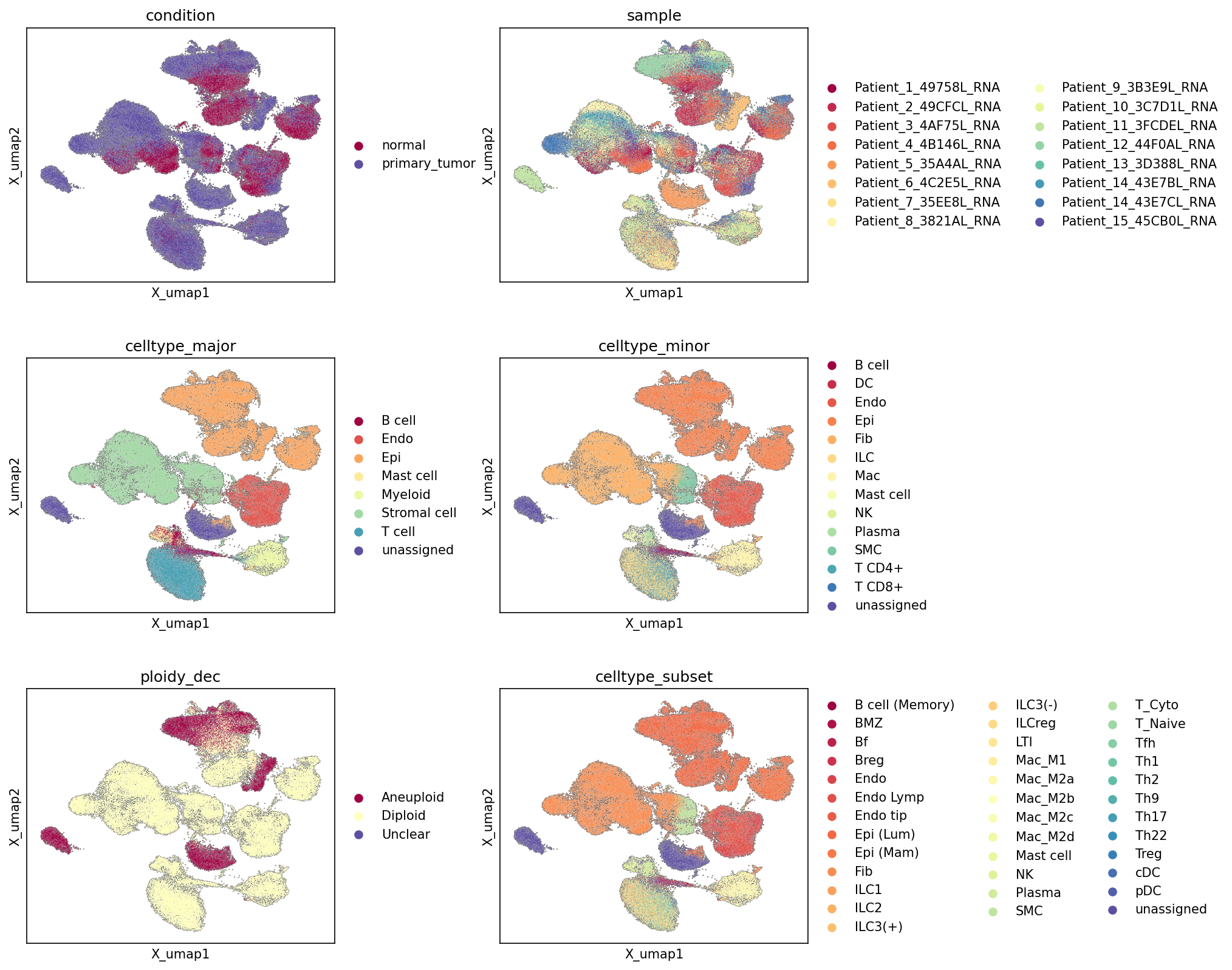
[Analysis Visualization Results]...
Analysis Overview
This analysis presents Uniform Manifold Approximation and Projection (UMAP) visualizations of single-cell RNA sequencing data from breast tissue, colored by various metadata features: condition (normal vs. primary tumor), sample, celltype_major, celltype_minor, ploidy_dec, and celltype_subset. These plots provide an overview of the dataset structure, cell type annotation quality, and the distribution of cells across different biological and technical covariates.
Visual Summary
Condition
The UMAP colored by condition shows a clear separation between normal (maroon) and primary_tumor (purple) cells. While some regions of the UMAP are mixed, indicating shared cell types or cell states between conditions, distinct clusters dominated by either normal or primary_tumor cells are evident. Notably, a large, well-defined cluster in the lower-right area of the UMAP is almost exclusively composed of primary_tumor cells, suggesting a population highly specific to the tumor microenvironment.
Sample
The sample plot displays a diverse distribution of individual samples across the UMAP. Most major cell clusters appear to be composed of cells from multiple patients, indicating good integration of data across different samples and suggesting that the observed clustering is driven by biological variation rather than strong batch effects. Some smaller, peripheral clusters might show enrichment for one or a few samples, which is common for rare cell populations or specific sample characteristics.
Celltype_major
This plot reveals distinct clustering according to broad cell types. Epithelial cell (orange) forms a large, somewhat diffuse cluster, often situated centrally or in regions with other structural cells. Stromal cell (light green) and Endothelial cell (red-orange) also form substantial clusters. Immune cell populations like T cell (cyan), Myeloid cell (light yellow), and B cell (dark red) form separate, well-defined clusters, indicating robust differentiation of these major lineages. A relatively small proportion of cells are labeled as unassigned (purple), scattered throughout the embedding.
Celltype_minor
The celltype_minor plot provides a more granular view of cell populations. Within the Epithelial cell major cluster, specific minor types are not explicitly broken out but are represented by a continued orange coloration, indicating the overall epithelial compartment. Fibroblast (light orange), Endothelial cell (red-orange), Macrophage (yellow), T cell CD8+ (dark blue), and T cell CD4+ (light blue) show more defined boundaries and separation within their respective major cell type compartments. Plasma cell (green) appears as a distinct, smaller cluster.
Ploidy_dec
The ploidy_dec plot highlights cells inferred as Aneuploid (maroon) and Diploid (yellow). A significant proportion of Aneuploid cells are concentrated within the large primary_tumor-specific cluster observed in the condition plot, and also in other smaller, specific regions. The majority of cells are Diploid and are broadly distributed across the embedding. A very small number of cells are labeled Unclear (purple) and are sparsely distributed. This strong spatial correlation between Aneuploid cells and primary_tumor condition suggests these aneuploid cells are likely the malignant epithelial cells.
Celltype_subset
This plot offers the finest resolution of cell types, showing numerous distinct subsets. For instance, different T cell subsets (e.g., T cell (Cytotoxic), T cell (Tfh), T cell (Treg)) are visible as sub-clusters within the broader T cell region. Macrophage subsets (Macrophage (M1), Macrophage (M2A), M2B, M2C, M2D) also show differential localization. Luminal epithelial cell and Mammary epithelial cell differentiate within the epithelial compartment. This level of annotation confirms the intricate cellular heterogeneity within the dataset.
Biological Interpretation
The UMAP visualizations provide critical insights into the cellular landscape of breast tissue and primary tumors:
- Tumor vs. Normal Segregation: The pronounced separation of primary_tumor and normal cells on the UMAP reflects significant global transcriptomic changes driven by the cancerous state. This indicates robust differential gene expression patterns exist between these conditions, making downstream analyses like differential expression and pathway enrichment highly informative for identifying tumor-specific biology.
- Malignant Cell Identification: Given that the Tumor origin celltype is Epithelial cell and aneuploidy is a hallmark of cancer, the strong co-localization of Aneuploid cells with primary_tumor regions, particularly within the large Epithelial cell cluster, provides strong evidence for the identification of the malignant epithelial cell population. This is a crucial finding for understanding tumor biology and progression.
- Heterogeneity of the Tumor Microenvironment: The presence and distinct clustering of various immune (T cells, B cells, Myeloid cells, ILCs, Mast cells, NK cells) and stromal (Fibroblasts, Endothelial cells, Smooth muscle cells) cell types in both normal and tumor contexts highlight the complex cellular composition of the breast tissue and the tumor microenvironment. The ability to resolve these cell types down to celltype_subset level allows for detailed investigation into their specific roles and interactions within the tumor.
- Annotation Quality and Data Structure: The clear and consistent clustering of cells by celltype_major, celltype_minor, and celltype_subset across the UMAP indicates high-quality cell type annotation. The hierarchical refinement from major to minor to subset annotations effectively captures the biological hierarchy of cell identity. The good mixing of sample origins within cell type clusters suggests successful integration of data from multiple patients, minimizing potential batch effects and ensuring that biological signals dominate the embedding structure.
Annotation Notes
The UMAP plots demonstrate generally high-quality annotations. Cells labeled as unassigned are minimal and scattered, suggesting that most cells have been confidently assigned to a known cell type. The clear separation of cell types at multiple granularities, and the meaningful co-localization of Aneuploid cells with the primary_tumor condition, reinforce the reliability of the cell type and ploidy annotations. This robust foundation is essential for subsequent in-depth analyses, such as cell-cell interaction studies or pathway analyses, by ensuring that comparisons are made between well-defined and biologically relevant cell populations.
2. Major Cell Type Score and Annotation Mapping on UMAP
[Analysis Visualization Results]...
Analysis Overview
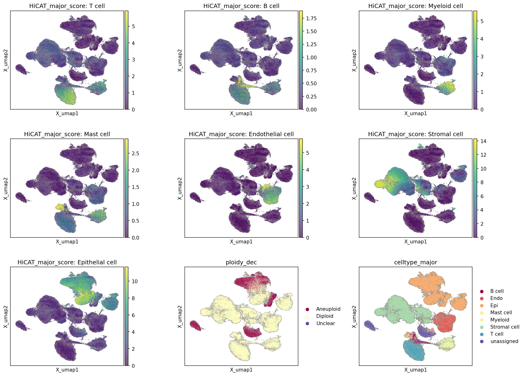
This analysis visualizes single-cell RNA sequencing data on a UMAP (Uniform Manifold Approximation and Projection) embedding. The plots display various major cell type scores, inferred by HiCAT, along with the inferred ploidy status and the pre-existing major cell type annotations. This allows for an assessment of cell type identification, the consistency between computational scoring and annotations, and the distribution of aneuploidy across the cellular landscape.
Visual Summary
The provided UMAP plots offer a comprehensive view of cell type distribution and characteristics within the dataset:
- HiCAT Major Cell Type Scores (T cell, B cell, Myeloid cell, Mast cell, Endothelial cell, Stromal cell, Epithelial cell): Each of these seven plots highlights distinct regions on the UMAP where the corresponding cell type score is high (indicated by warmer colors, typically yellow). These high-scoring regions largely segregate into discrete clusters, suggesting distinct transcriptional profiles for each major cell type.
- T cells are predominantly found in a cluster in the bottom-right and a smaller cluster in the top-left.
- B cells show a distinct, smaller cluster in the bottom-middle.
- Myeloid cells are concentrated in a cluster in the bottom-right, distinct from T cells.
- Mast cells form a small, localized cluster in the bottom-middle, often adjacent to B cells.
- Endothelial cells define a cluster in the mid-right region.
- Stromal cells occupy a large, somewhat diffuse region spanning the left side and extending towards the center of the UMAP.
- Epithelial cells form a prominent, large cluster in the top-middle/top-right region.
- Ploidy Status (ploidy_dec): This plot categorizes cells into 'Aneuploid' (dark red), 'Diploid' (light yellow), and 'Unclear' (dark blue). A significant cluster of Aneuploid cells is prominently located in the top-middle/top-right region of the UMAP. The vast majority of cells in other clusters are labeled as Diploid.
- Major Cell Type Annotation (celltype_major): This plot displays the pre-assigned celltype_major labels for each cell using distinct colors. This serves as a reference for validating the HiCAT major cell type scores and the overall clustering structure.
- The visual distribution of celltype_major annotations for B cell, Endothelial cell, Epithelial cell, Mast cell, Myeloid cell, Stromal cell, and T cell corresponds remarkably well with the high-score regions observed in their respective HiCAT major cell type score plots. This indicates high consistency between the HiCAT scoring and the existing annotations.
- The unassigned cells are scattered across the UMAP, with some forming small, less distinct clusters.
Biological Interpretation
- Robust Cell Type Identification: The strong spatial segregation of different HiCAT major cell type scores into distinct UMAP clusters, which are highly concordant with the celltype_major annotations, suggests a robust and consistent cell type identification process. This indicates that the major cell populations within the breast tissue single-cell dataset are well-defined by their transcriptomic profiles.
- Identification of Tumor Cells via Aneuploidy: A striking observation is the clear co-localization of the Epithelial cell cluster (as shown by both HiCAT score and celltype_major annotation) with the Aneuploid cell population. Given that the Tumor origin celltype is specified as 'Epithelial cell' and the tissue is 'Breast' with 'primary_tumor' and 'normal' conditions, this strong overlap strongly indicates that the Aneuploid cells represent the malignant epithelial cells of the breast tumor. Aneuploidy, the presence of an abnormal number of chromosomes, is a hallmark of cancer cells and is frequently observed in breast carcinoma [1].
[1] PubMed search for "aneuploidy breast cancer"
- Tumor Microenvironment Composition: The remaining clusters, predominantly composed of Diploid cells, represent the tumor microenvironment (TME) or normal breast tissue components. These include:
- Stromal cells: A large, diverse population potentially encompassing fibroblasts, smooth muscle cells, and adipocytes that contribute to the structural and functional support of the tissue, both normal and cancerous.
- Immune cells: T cells, B cells, Myeloid cells (macrophages, dendritic cells), and Mast cells are all key players in the immune response within the TME, influencing tumor progression and response to therapy [2].
- Endothelial cells: These form the vasculature, crucial for oxygen and nutrient supply to both tumor and normal tissue.
- The spatial arrangement on the UMAP hints at potential relationships or shared microenvironments between these cell types. For example, stromal cells often interact closely with epithelial cells in both normal and tumor contexts.
- Annotation Quality: The high degree of concordance between the HiCAT cell type scores and the celltype_major annotations validates the quality of the existing cell type assignments. This consistency strengthens confidence in downstream analyses that rely on these cell type labels.
Annotation Notes
The UMAP plots demonstrate excellent segregation of major cell types, with both the computational HiCAT scores and the celltype_major annotations defining clear, distinct clusters. The crucial finding of aneuploid cells coinciding perfectly with the epithelial cell population in the tumor context provides strong evidence for accurate identification of malignant cells. The overall embedding structure is well-defined, and the cell identity annotations appear robust.
3. Overall Celltype_subset Marker Expression Dot Plot Analysis
[Analysis Visualization Results]...
Analysis Overview
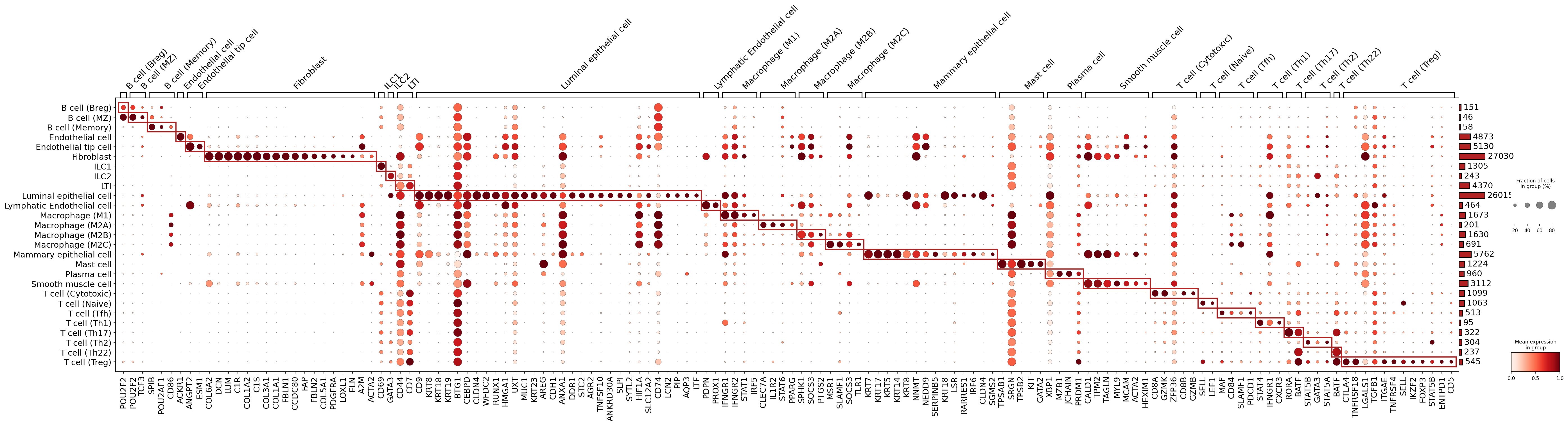
This analysis presents a dot plot illustrating the expression patterns of marker genes across various celltype_subset populations identified in the single-cell RNA sequencing (scRNA-seq) dataset. The primary goal of this visualization is to validate the assigned cell type identities by assessing whether each celltype_subset expresses known canonical marker genes. The plot displays both the mean expression level (color intensity) and the fraction of cells expressing each gene (dot size) within each cell group. The red boxes highlight marker genes most enriched within specific cell type subsets, aiding in identity confirmation.
Visual Summary
The dot plot is structured with celltype_subset annotations on the y-axis and identified marker genes on the x-axis.
- Dot Size: The size of each dot corresponds to the percentage of cells within a given celltype_subset that express the specific marker gene. Larger dots indicate a higher fraction of expressing cells.
- Dot Color: The intensity of the red color in each dot represents the mean expression level of the marker gene in that celltype_subset. Darker red indicates higher mean expression.
- Grouped Markers: Genes are grouped by the celltype_subset they characterize, and these groups are visually delineated by red boxes, making it easy to identify cell-type-specific gene signatures.
- Cell Counts: A bar chart on the right side indicates the total number of cells contributing to each celltype_subset group. Most groups appear to have a sufficient number of cells for robust analysis (e.g., Fibroblast ~27k cells, Luminal epithelial cell ~26k cells).
Overall, a clear diagonal pattern of strong red, large dots within the red boxes is observed, indicating distinct and specific marker gene expression for most celltype_subset populations. This pattern suggests that the current celltype_subset annotations are well-supported by their respective gene expression profiles.
Biological Interpretation
The marker gene expression patterns strongly support the assigned celltype_subset identities, validating the annotation quality of the dataset.
- B cells: Subsets like B cell (Breg), B cell (MZ), and B cell (Memory) show strong expression of classical B cell markers such as *POU2F2* (OCT2, a B-cell transcription factor), *POU2AF1* (OBF-1, B-cell specific coactivator), *TCF3*, and *SPIB*. Plasma cells, differentiated B cells, are also distinct, expressing *XBP1*, *MZB1*, *JCHAIN*, and *PRDM1*, consistent with their role in antibody production.
- POU2F2 GeneCards
- XBP1 GeneCards
- Endothelial cells: Both Endothelial cell and Endothelial tip cell populations are clearly identified by markers like *ACKR1* (DARC), *ANGPT2* (Angiopoietin-2), *ESM1*, *CD34*, *PECAM1* (CD31), *FLT1*, and *SELE*. Lymphatic Endothelial cells express *PROX1* and *PDPN*, which are canonical lymphatic endothelial markers.
- PECAM1 GeneCards
- PROX1 GeneCards
- Fibroblasts: These cells exhibit high expression of extracellular matrix (ECM) related genes such as *DCN* (Decorin), *LUM* (Lumican), *COL1A1*, *COL1A2*, *COL3A1*, *COL5A1*, *FBLN1*, *FAP* (Fibroblast Activation Protein), and *PDGFRA*, confirming their identity as connective tissue cells.
- DCN GeneCards
- FAP GeneCards
- Innate Lymphoid Cells (ILC): The various ILC subsets (ILC1, ILC2, LTI) show distinct marker expression. ILC2 cells strongly express *GATA3*, a key transcription factor for type 2 immunity. LTI (Lymphoid Tissue inducer) cells show expression of *TNFSF10*.
- GATA3 GeneCards
- Epithelial cells: Luminal epithelial cells and Mammary epithelial cells are clearly marked by a panel of keratins (*KRT7, KRT8, KRT14, KRT17, KRT18, KRT19*), along with *MUC1* and *CDH1* (E-cadherin), which are characteristic of epithelial lineages in the breast.
- KRT18 GeneCards
- CDH1 GeneCards
- Macrophages: Different macrophage subsets (M1, M2A, M2B, M2C, M2D) show common macrophage markers like *CD68* and *MSR1*. Specific polarization states are hinted at by markers like *CD86* (more M1-associated) and *CLEC7A* (Dectin-1).
- CD68 GeneCards
- Mast cells: These cells are strongly characterized by *KIT* (CD117), *TPSAB1* (Tryptase alpha/beta 1), and *TPSB2* (Tryptase beta 2), which are well-known mast cell-specific genes.
- KIT GeneCards
- Smooth muscle cells: Marked by *ACTA2* (alpha-smooth muscle actin), *MYL9*, and *TAGLN*, consistent with their contractile function.
- ACTA2 GeneCards
- T cells: Various T cell subsets (Cytotoxic, Naive, Tfh, Th1, Th17, Th2, Th22, Treg) display highly specific marker profiles.
- T cell (Cytotoxic): Characterized by *CD8A*, *GZMK*, *GZMB*, consistent with their killer function.
- T cell (Naive): Express *SELL* (CD62L) and *LEF1*, indicating their naive state.
- T cell (Tfh): Show markers like *PDCD1* (PD-1), *ICOS*, and *CXCR5*, which are crucial for B cell help.
- T cell (Th1): Express *STAT1* and IFN-gamma receptor components (*IFNGR1*, *IFNGR2*), aligning with their role in cell-mediated immunity.
- T cell (Th17): Characterized by *RORC*, the master regulator of Th17 differentiation.
- T cell (Treg): Strongly express *FOXP3*, *CTLA4*, *TNFRSF18* (GITR), and *ENTPD1* (CD39), confirming their immunosuppressive phenotype.
- CD8A GeneCards
- FOXP3 GeneCards
- Dendritic cells: DC (Classical) show expression of *CD1C*, a common marker for conventional DCs.
- CD1C GeneCards
Annotation Notes
The dot plot demonstrates excellent concordance between the assigned celltype_subset labels and their respective marker gene expression profiles. The specificity and enrichment of markers within their expected cell types are high, with minimal off-target expression observed for most key markers. The number of cells per group is generally robust, providing confidence in the observed expression patterns. This analysis provides strong evidence for the high quality and accuracy of the cell type annotations in this AnnData object. The clear segregation of markers confirms that the clustering and annotation workflow successfully captured biologically distinct cell populations.
4. Analysis of Copy Number Variations in Tumor-Origin and Unassigned Cells
[Analysis Visualization Results]...
Analysis Overview
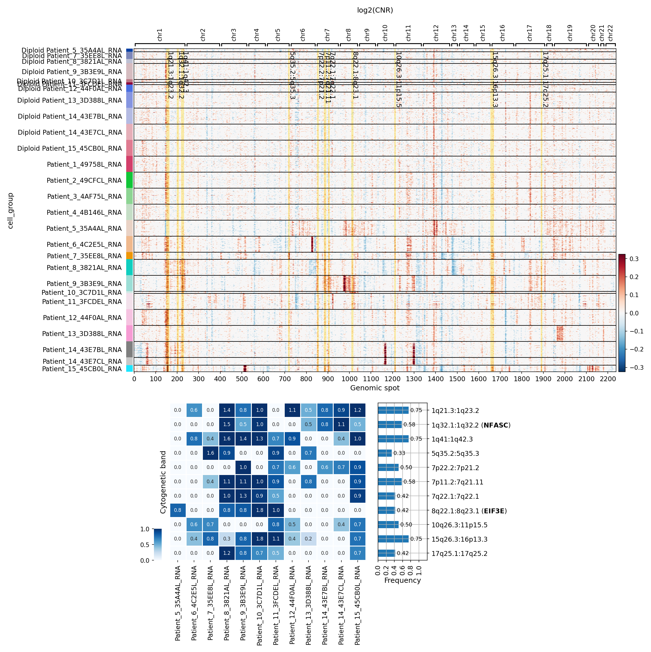
This analysis investigates copy number variations (CNVs) in cells identified as 'Epithelial cell' (the tumor-origin cell type in breast tissue) and 'unassigned' cells. The CNV estimates, obtained from single-cell RNA-seq data, are visualized as a heatmap grouped by individual samples, distinguishing between diploid and aneuploid cell populations. A summary of significantly amplified cytogenetic regions across samples is also provided to highlight recurrent genomic alterations.
Visual Summary
The CNV heatmap displays log2(CNR) values across genomic spots for the targeted cell populations. Amplifications are shown in red, and deletions in blue.
- Ploidy-driven Grouping: The heatmap is distinctly divided into two main sections. The upper section comprises cells labeled with a "Diploid" prefix (e.g., "Diploid Patient_5_35A4AL_RNA"), which generally exhibit minimal or no significant CNVs, appearing largely blue or white, consistent with a diploid genomic state.
- Aneuploid Samples: The lower section, labeled without the "Diploid" prefix (e.g., "Patient_1_497F8L_RNA"), shows extensive and widespread CNVs, characterized by prominent red (amplification) and blue (deletion) bands across many chromosomes. This pattern is indicative of aneuploidy and genomic instability, typical of malignant cells.
- Sample-Specific CNV Profiles: Within the aneuploid group, individual samples demonstrate distinct CNV patterns, though some recurrent alterations are visible. For example, Patient_7, Patient_9, Patient_10, Patient_11, Patient_14, and Patient_15 show particularly prominent amplification events, especially on chromosome 1, 8, and other regions.
- Recurrent Amplifications: The accompanying summary heatmap and bar plot highlight frequently amplified cytogenetic bands across the aneuploid samples. The most frequently amplified regions, with a frequency greater than 75% across samples, include:
1q21.3:1q23.2
1q32.1:1q32.2 (containing the *NFASC* gene)
1q41:1q42.3
8q22.1:8q23.1 (containing the *EIF3E* gene)
Other frequently amplified regions (frequency > 50%) include various segments on chromosomes 5, 7, 10, 15, and 17.
Biological Interpretation
The striking difference in CNV landscapes between the "Diploid" and "Aneuploid" patient groups among the 'Epithelial cell' and 'unassigned' populations underscores the hallmark of genomic instability in tumor cells.
- Malignant Nature of Aneuploid Cells: Given that 'Epithelial cell' is designated as the tumor-origin cell type in breast tissue, the cells from "Patient_X" (non-Diploid labeled samples) showing extensive CNVs are highly likely to be malignant epithelial cells. The 'unassigned' cells, if they also show extensive CNVs in these aneuploid samples, might represent poorly annotated tumor cells or a mixture of cell types with similar genomic instability.
- Recurrent Amplifications in Breast Cancer: The identification of recurrent amplifications points to regions of the genome that are preferentially selected for gain during tumor evolution.
- Chromosome 1q Amplifications: The high frequency of amplifications on the long arm of chromosome 1 (1q21.3:1q23.2, 1q32.1:1q32.2, 1q41:1q42.3) is a common finding in many cancers, including breast cancer. This region harbors several oncogenes and genes involved in cell proliferation and survival. For instance, *NFASC* (Neurofascin) at 1q32.1 has been implicated in cell migration and invasion in various cancers, and its amplification might contribute to an aggressive phenotype PubMed Search: NFASC cancer amplification.
- Chromosome 8q Amplifications: The recurrent amplification of 8q22.1:8q23.1, including the *EIF3E* gene, is also significant. The 8q arm frequently shows gains in breast cancer. *EIF3E* (Eukaryotic Translation Initiation Factor 3 Subunit E) is part of a complex crucial for protein synthesis. Upregulation of eIF3 components, including *EIF3E*, can promote cell proliferation and survival, making it a potential oncogene when amplified GeneCards: EIF3E.
- Genomic Instability as a Driver: The widespread nature of CNVs across multiple chromosomes in the aneuploid samples highlights the high degree of genomic instability inherent to these tumor cells, which drives tumor heterogeneity and evolution.
Clinical or Translational Implications
- Prognostic and Predictive Markers: Recurrent CNVs, such as the amplifications on 1q and 8q, can serve as potential prognostic markers or indicators of disease aggressiveness in breast cancer. Further research could validate these regions as biomarkers for patient stratification.
- Therapeutic Vulnerabilities: Genes located within frequently amplified regions, like *NFASC* and *EIF3E*, may represent "driver" oncogenes whose increased copy number leads to overexpression and dependency. Such genes could be explored as potential therapeutic targets, especially if their activation is critical for tumor maintenance.
- Understanding Tumor Evolution: Characterizing the CNV landscape provides insight into the evolutionary trajectories of breast tumors, showing how different clones within a tumor acquire specific genomic aberrations. This understanding can aid in predicting resistance mechanisms and designing more effective combination therapies.
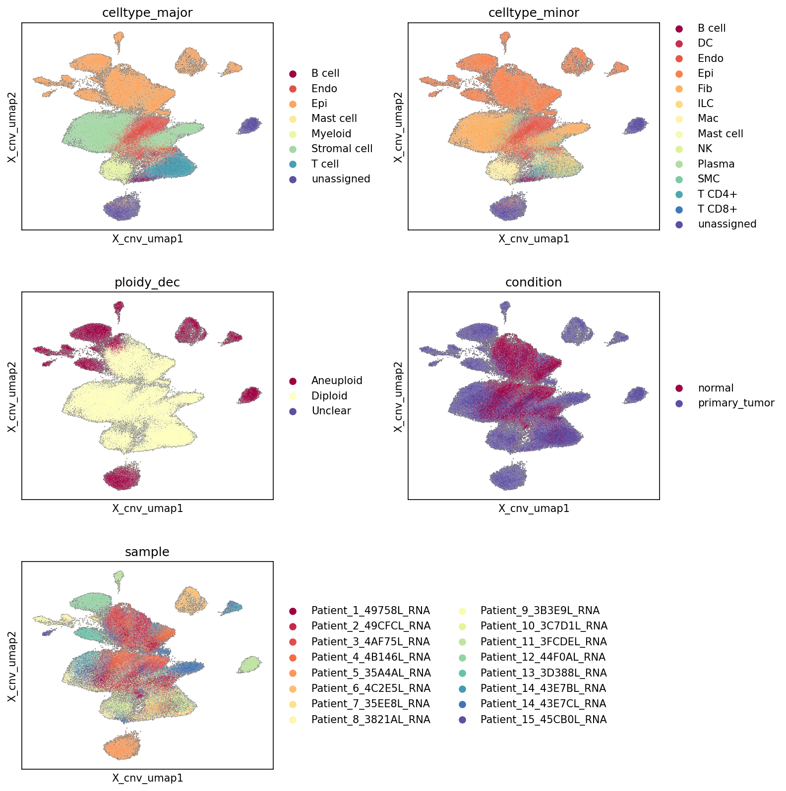
5. CNV-based UMAP Visualization of Breast Tissue Single-cell RNA-seq Data
[Analysis Visualization Results]...
Analysis Overview
This analysis presents a Uniform Manifold Approximation and Projection (UMAP) visualization generated specifically from Copy Number Variation (CNV) estimates for single cells from breast tissue. This approach allows for the unsupervised clustering of cells based on their genomic alterations, rather than gene expression patterns. The UMAP plots are colored by major cell type, minor cell type, ploidy declaration (ploidy_dec), condition (primary tumor vs. normal), and individual sample to reveal underlying structural relationships and heterogeneity driven by CNV profiles.
Visual Summary
Embedding Structure and Ploidy
The UMAP clearly segregates cells into distinct clusters based on their CNV profiles. A prominent observation is the robust separation based on ploidy:
- Diploid cells (yellow in ploidy_dec plot) form a large, cohesive central mass, representing cells with stable, non-aneuploid genomes. These are likely non-malignant cells.
- Aneuploid cells (dark red in ploidy_dec plot) occupy several distinct, spatially separated clusters, notably a large cluster on the left and smaller, more diffuse clusters elsewhere. This segregation indicates that aneuploid cells possess unique and diverse genomic alteration patterns that differentiate them from diploid cells and also from each other.
- Cells labeled as "Unclear" ploidy are scattered, often found at the interfaces between diploid and aneuploid regions or in less defined clusters.
Cell Type Distribution
- Major Cell Types: The large central cluster, predominantly diploid, is enriched with various stromal and immune cells including Stromal cells, Endothelial cells, Myeloid cells, T cells, B cells, and Mast cells.
- Crucially, Epithelial cells (Epi), identified as the tumor origin cell type, show a strong enrichment within the distinct, left-most aneuploid cluster. They also appear to have some representation in the central diploid region, which might correspond to normal epithelial cells or cells with less pronounced aneuploidy.
- Minor Cell Types: This granularity further supports the major cell type observations. Fibroblasts, Macrophages, T cell CD4+, T cell CD8+, Plasma cells, and Endothelial cells largely constitute the diploid populations. Luminal epithelial cells and other epithelial subsets likely comprise the distinct aneuploid clusters.
Condition and Sample Distribution
Condition:
- Normal samples (dark red in condition plot) are almost exclusively confined to the large, central diploid cluster, as expected for healthy tissue.
- Primary tumor samples (purple in condition plot) are broadly distributed across the UMAP. They heavily overlap with the central diploid population, indicating the presence of tumor microenvironment (TME) cells (immune, stromal, endothelial) within the tumor tissue. More importantly, primary tumor cells are the sole contributors to the distinct aneuploid clusters, directly associating aneuploidy with the tumor condition.
- Sample: The sample plot highlights significant inter-patient heterogeneity. While the central diploid clusters show mixed contributions from various patients, the aneuploid clusters are often dominated by cells from specific patients. For example, the large left aneuploid cluster is primarily formed by cells from patients like Patient_1, Patient_2, Patient_3, Patient_4, Patient_5, suggesting unique or shared CNV profiles among these tumor cells. Other patients, such as Patient_14 and Patient_15, contribute to other distinct aneuploid clusters, illustrating the diverse genomic landscape across individual tumors.
Biological Interpretation
The CNV-driven UMAP provides compelling evidence for genomic instability as a primary driver of cellular heterogeneity in breast cancer.
- Malignant Cell Identification: The strong co-localization of Aneuploid cells with Epithelial cells from primary_tumor samples strongly identifies these cells as the malignant tumor cells. This separation from the predominantly Diploid stromal and immune cells reinforces the genomic basis of tumor identity.
- Tumor Microenvironment (TME) Composition: The presence of a substantial Diploid cell population (comprising stromal, immune, and endothelial cells) within the primary_tumor samples, co-clustering with normal cells, accurately reflects the complex cellular ecosystem of the TME, which consists of non-malignant host cells.
- Genomic Heterogeneity within Tumors: The observation that Aneuploid cells from different samples form distinct clusters on the CNV-UMAP suggests significant inter-patient genomic heterogeneity in breast cancer. This implies that while all tumor cells may share a general aneuploid status, the specific chromosomal gains and losses can vary widely between individuals, potentially influencing tumor biology and treatment response.
- Robustness of Ploidy Inference: The clear separation of Aneuploid from Diploid cells, especially given that the UMAP embedding itself is based on CNV estimates, validates the accuracy and biological relevance of the ploidy_dec annotation. Cells labeled as Unclear often reside in transitional or ambiguous genomic states, which might represent cells with low levels of aneuploidy, partial CNVs, or technical limitations in their CNV estimation.
Annotation Notes
- The celltype_major, celltype_minor, condition, and sample annotations align exceptionally well with the ploidy_dec states in the CNV-based UMAP. This provides strong confidence in the cell type assignments and the distinction between normal and tumor conditions based on genomic integrity.
- The clustering of Epithelial cells within the Aneuploid regions from primary_tumor samples is consistent with the Tumor origin celltype context, validating the identification of malignant cells.
- The clear separation of normal tissue components from the tumor-specific aneuploid populations underscores the utility of CNV analysis in delineating tumor cells from the surrounding microenvironment in single-cell datasets.
6. Minor Cell Type Population Analysis in Breast Tissue
[Analysis Visualization Results]...
Analysis Overview
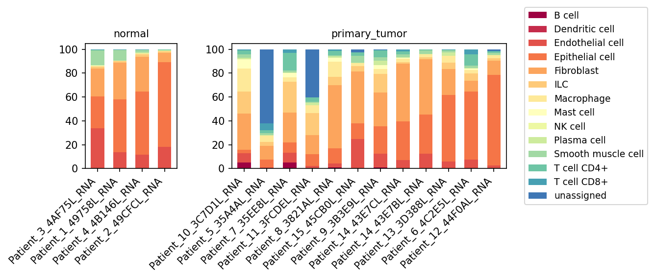
This analysis provides a population bar plot illustrating the relative proportions of different minor cell types across individual samples from both normal breast tissue and primary breast tumors. This visualization is crucial for understanding the cellular composition of the breast tissue microenvironment in health and disease, highlighting shifts associated with tumor development.
Visual Summary
The stacked bar plots display the percentage composition of various minor cell types for each sample, grouped by condition (normal vs. primary_tumor).
Normal Tissue Composition:
- Normal breast tissue samples show a relatively consistent cellular composition.
- Epithelial cells (orange) and Fibroblasts (light orange) constitute the largest fractions, collectively making up a substantial portion of the normal tissue.
- Endothelial cells (red) are also consistently present in considerable proportions.
- Immune cells such as Macrophages (yellow), T cells (CD4+ and CD8+) (teal shades), and other minor populations are present but generally at lower frequencies.
- The "unassigned" category is minimal in normal samples.
Primary Tumor Tissue Composition:
- Primary tumor samples exhibit greater heterogeneity in cell type composition compared to normal samples.
- Epithelial cells (orange) generally remain a dominant population, consistent with their role as the cell of origin for breast cancer. However, their proportions can vary significantly between tumor samples.
- Fibroblasts (light orange) are also consistently abundant in tumor samples, often comparable to or even slightly higher than in normal tissue, indicating a robust stromal component.
- A striking feature in tumor samples is the increased and variable presence of "unassigned" cells (dark blue) in several samples (e.g., Patient_10, Patient_7, Patient_11, Patient_8). This suggests that a significant fraction of cells in these tumors may not fit the established minor cell type annotations or represent novel/altered cell states.
- The immune cell landscape is more complex and variable in tumors. While Macrophages (yellow) appear more prominent in some tumor samples than in normal tissue, other immune cells like T cells show fluctuating proportions.
- Endothelial cells (red) are present, reflecting tumor vascularization.
Biological Interpretation
The observed shifts in minor cell type populations between normal and primary tumor breast tissue provide critical insights into the tumor microenvironment (TME) dynamics:
- Dominance of Epithelial and Stromal Cells: The consistent high proportion of Epithelial cells in both normal and tumor tissue aligns with the epithelial origin of most breast cancers. The sustained high presence of Fibroblasts (often referred to as Cancer-Associated Fibroblasts, CAFs, in tumors) highlights their crucial role in tumor progression, extracellular matrix remodeling, and creation of a pro-tumorigenic niche https://pubmed.ncbi.nlm.nih.gov/30678683/.
- Immune Cell Infiltration and Heterogeneity:
- The variable presence of immune cells, particularly Macrophages, T cells (CD4+ and CD8+), and other lymphoid populations (B cells, NK cells, Dendritic cells), underscores the immune-edited nature of the tumor microenvironment. Macrophages, often polarized to M2-like phenotypes in tumors, are known to promote angiogenesis, immune suppression, and metastasis https://pubmed.ncbi.nlm.nih.gov/31105260/.
- The fluctuating levels of T cells suggest inter-patient variability in immune response and infiltration, which can be critical for prognosis and response to immunotherapies.
- Significant "Unassigned" Population in Tumors: The notable increase in "unassigned" cells in several primary tumor samples is a key observation. This could represent:
- Tumor-specific cell states: Highly aberrant or dedifferentiated tumor cells that deviate significantly from their normal epithelial counterparts, making them difficult to annotate with standard references.
- Novel stromal or immune subtypes: Unique populations of stromal or immune cells that emerge or are significantly altered within the TME and are not captured by existing minor cell type definitions.
- Technical artifacts or low-quality cells: While less likely to be consistently high across multiple samples without annotation, it's a possibility.
Further investigation into these "unassigned" cells is warranted to characterize their identity and potential functional roles in tumor biology.
- Tissue Specificity: This analysis is highly relevant to breast cancer, where the interplay between epithelial tumor cells, fibroblasts, and infiltrating immune cells is well-documented in driving disease progression and therapeutic resistance.
Clinical or Translational Implications
- Biomarker Discovery: The distinct cellular compositions, especially the presence of certain immune or stromal cell types and the "unassigned" population, could serve as prognostic or predictive biomarkers for breast cancer outcomes or response to specific treatments.
- Therapeutic Targeting: Identifying cell types that are significantly enriched in primary tumors (e.g., CAFs, specific macrophage subtypes) could point to novel therapeutic targets. For instance, strategies aimed at depleting CAFs or re-educating tumor-associated macrophages are under investigation in cancer therapy.
- Understanding Treatment Resistance: The heterogeneity in cell type composition across different tumor samples suggests that patients may respond differently to therapies based on their unique TME. For example, tumors with higher infiltration of suppressive immune cells might benefit from immunotherapy, while those dominated by fibroblasts might require stromal-targeting agents.
- Improving Cell Annotation: The substantial "unassigned" population highlights the ongoing need for refined annotation strategies in single-cell cancer atlases. Characterizing these cells could unveil novel malignant or TME-resident cell states with unique biological and clinical implications.
7. T Cell Subpopulation Analysis in Normal vs. Primary Breast Tumor Tissue
[Analysis Visualization Results]...
Analysis Overview
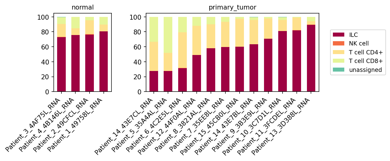
This analysis visualizes the proportional distribution of minor cell types within the major cell type category "T cell" across normal and primary breast tumor samples. The plot_celltype_population tool was used to break down the 'T cell' major population into its celltype_minor constituents, which include T cell CD4+, T cell CD8+, ILC, NK cell, and unassigned populations. This comparison aims to highlight shifts in immune cell composition between healthy and cancerous breast tissue microenvironments.
Visual Summary
The stacked bar plot displays the relative proportions of T cell subsets and related lymphoid cells per sample, grouped by condition (normal vs. primary_tumor).
- Normal Condition: In the normal breast tissue samples (left panel), Innate Lymphoid Cells (ILCs, dark red) are the predominant population, constituting a large majority (typically >70%) of the cells within this broader "T cell" major category. T cell CD4+ (light yellow) and T cell CD8+ (light green) are present but in smaller, relatively consistent proportions across normal samples. NK cells (orange) and unassigned cells (teal) are negligible.
- Primary Tumor Condition: The primary tumor samples (right panel) exhibit significantly higher heterogeneity in cell subpopulation proportions compared to normal samples.
- Some tumor samples (e.g., Patient_14_43E7CL_RNA, Patient_5_35A4AL_RNA) show a marked decrease in the relative proportion of ILCs, with a concomitant increase in T cell CD4+ and T cell CD8+ populations. In these samples, T cell CD4+ often becomes the most abundant population, followed by ILCs and then T cell CD8+.
- Other tumor samples (e.g., Patient_11_3FCDEL_RNA, Patient_13_3D388L_RNA) maintain a high proportion of ILCs, similar to or even exceeding normal samples, while still showing varied but generally lower contributions from CD4+ and CD8+ T cells.
- The presence of NK cells and unassigned cells remains consistently low across all tumor samples.
Biological Interpretation
The analysis reveals distinct lymphoid cell compositions in normal breast tissue versus primary breast tumors, with significant heterogeneity within the tumor microenvironment.
- Broad Definition of "T cell" Major Category: It's important to note that within this dataset, the celltype_major category "T cell" appears to encompass a broader lymphoid compartment, as its celltype_minor breakdown includes not only conventional T cells (CD4+, CD8+) but also Innate Lymphoid Cells (ILCs) and Natural Killer (NK) cells. This implies a hierarchical classification where these distinct lymphocyte populations are initially grouped under a common "T cell" major label before more granular annotation.
- ILC Dominance in Normal Breast Homeostasis: The consistent dominance of ILCs in normal breast tissue suggests their crucial role in maintaining immune surveillance and tissue homeostasis in healthy mammary glands. ILCs are key regulators of inflammation, tissue repair, and immune responses at mucosal and barrier surfaces, which would include the breast. Specific ILC subsets (ILC1, ILC2, ILC3) have diverse functions, and their overall abundance indicates a highly active innate lymphoid immune component.
- T Cell Infiltration and Immunosuppression in Tumors: The observed increase in CD4+ and CD8+ T cell proportions in a subset of primary tumor samples indicates active immune cell infiltration into the tumor microenvironment (TME).
- CD8+ T cells are cytotoxic T lymphocytes (CTLs) that play a central role in anti-tumor immunity. An increased presence is often associated with a more "immune-inflamed" tumor phenotype.
- CD4+ T cells can act as helper cells (e.g., Th1, Th17) or regulatory cells (Tregs) that modulate anti-tumor responses. Their specific functional subtype would need further investigation.
- Conversely, tumor samples where ILCs remain highly dominant and T cells are relatively sparse might represent an "immune-desert" or "immune-excluded" phenotype, where anti-tumor T cell responses are limited or suppressed. This could be driven by various mechanisms within the TME preventing T cell infiltration or activation.
- Heterogeneity of the Breast Cancer Immune Microenvironment: The marked variability in lymphoid cell composition among primary tumor samples underscores the profound heterogeneity of the breast cancer immune microenvironment. This suggests that different tumors might engage with the immune system in diverse ways, impacting their progression and responsiveness to immunotherapies.
Clinical or Translational Implications
The findings have several clinical and translational implications for breast cancer:
- Prognostic and Predictive Biomarkers: The proportions of ILCs, CD4+ T cells, and CD8+ T cells within the tumor microenvironment could serve as valuable prognostic or predictive biomarkers for breast cancer patients. Tumors with higher CD8+ T cell infiltration might be more responsive to immune checkpoint inhibitors (e.g., PD-1/PD-L1 blockade) https://pubmed.ncbi.nlm.nih.gov/33926668/.
- Targeting ILCs: The high baseline presence of ILCs in normal tissue, and their variable presence in tumors, suggests that understanding their specific roles (e.g., pro-tumorigenic vs. anti-tumorigenic) in breast cancer could open new therapeutic avenues. ILC2s, for example, have been implicated in promoting tumor growth in some contexts https://pubmed.ncbi.nlm.nih.gov/34293843/.
- Personalized Immunotherapy: The pronounced inter-tumor heterogeneity highlights the need for personalized approaches to immunotherapy. Comprehensive immune profiling of individual tumors, like this subpopulation analysis, could help stratify patients and guide the selection of appropriate treatment strategies.
- Immune Hot vs. Cold Tumors: The varying degrees of T cell infiltration suggest the existence of "immune-hot" (more T cells) and "immune-cold" (fewer T cells, potentially more ILCs) breast tumors. Strategies to convert "cold" tumors to "hot" ones, such as oncolytic viruses or specific chemotherapies, are areas of active research https://pubmed.ncbi.nlm.nih.gov/32959648/.
8. Differential Proportions of T Cell and ILC Subsets in Breast Primary Tumors
[Analysis Visualization Results]...
Analysis Overview
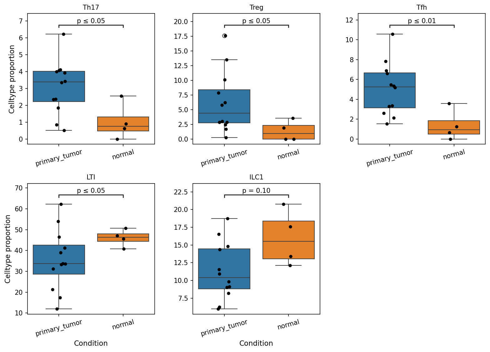
This analysis investigates the proportional differences of specific T cell and innate lymphoid cell (ILC) subsets within breast tissue, comparing primary tumor samples against normal tissue samples. The aim is to identify immune cell populations that are significantly altered in the tumor microenvironment, which can provide insights into tumor immunology and potential therapeutic targets. The plot_box_for_celltype_population_with_signif_difference tool was utilized to visualize these differences, specifically focusing on subsets within the 'T cell' major category and related ILCs, and highlighting those with a p-value less than or equal to 0.1.
Visual Summary
The box plots display the cell type proportions for five immune cell subsets: Th17, Treg, Tfh, LTI, and ILC1, comparing primary_tumor (blue boxes) and normal (orange boxes) conditions.
- Th17 cells: Exhibit a significantly higher median proportion in primary_tumor samples compared to normal samples (p ≤ 0.05).
- Treg cells: Show a notably higher median proportion in primary_tumor samples than in normal samples (p ≤ 0.05), with a wider spread in the tumor condition.
- Tfh cells: Are significantly more abundant in primary_tumor samples compared to normal samples (p ≤ 0.01), indicating a highly significant increase.
- LTI cells: Display a significantly lower median proportion in primary_tumor samples compared to normal samples (p ≤ 0.05), suggesting a depletion in the tumor microenvironment.
- ILC1 cells: While not meeting the conventional p < 0.05 threshold, show a trend towards a lower median proportion in primary_tumor samples compared to normal samples (p = 0.10).
Biological Interpretation
The observed alterations in the proportions of these specific T cell and ILC subsets in the breast primary tumor microenvironment provide critical insights into the immune landscape of breast cancer.
- Increased Immunosuppressive and Pro-inflammatory T cells (Treg, Th17, Tfh):
- Regulatory T cells (Treg): The significant enrichment of Tregs in primary tumors is a common hallmark of cancer. Tregs play a crucial role in suppressing anti-tumor immune responses, thereby contributing to immune evasion and tumor progression. Their high numbers can inhibit the activity of effector T cells (like cytotoxic T lymphocytes), natural killer cells, and antigen-presenting cells, fostering an immunosuppressive environment.
- T helper 17 (Th17) cells: Th17 cells are often implicated in both pro-tumorigenic and anti-tumorigenic roles, depending on the specific cancer context and cytokine milieu. In breast cancer, an increased presence of Th17 cells can be associated with chronic inflammation and can promote angiogenesis and tumor growth, especially in certain subtypes PubMed search for Th17 breast cancer. Their elevated proportion here could indicate a shift towards a pro-inflammatory state that supports tumor progression.
- T follicular helper (Tfh) cells: The significant increase in Tfh cells in primary tumors suggests an active B cell-mediated immune response within the tumor microenvironment or tumor-draining lymph nodes. Tfh cells are essential for germinal center formation and providing help to B cells for antibody production and memory B cell differentiation UniProt: CD40LG, BCL6. While B cell responses can be anti-tumor, they can also contribute to tumor growth or immune evasion through various mechanisms, making the functional outcome of increased Tfh cells context-dependent.
- Decreased Innate Lymphoid Cells (LTI, ILC1):
- Lymphoid Tissue Inducer (LTI) cells: LTI cells, a subset of ILC3s, are crucial for the development and maintenance of secondary lymphoid organs and tertiary lymphoid structures (TLS) GeneCards: RORC. The significant decrease of LTI cells in primary tumors suggests a potential disruption in the formation or maintenance of immune niches. TLS can either promote anti-tumor immunity or serve as sites for pro-tumorigenic immune responses; thus, their reduction could impact the overall immune organization within the tumor.
- ILC1 cells: ILC1s are innate counterparts to Th1 cells, producing IFN-gamma and TNF-alpha, which are critical for anti-tumor immunity against intracellular pathogens and cancer cells PubMed search for ILC1 anti-tumor immunity. The observed trend towards decreased ILC1s in tumors might indicate a weakened innate anti-tumor surveillance mechanism, allowing for tumor progression.
These shifts collectively paint a picture of an altered immune microenvironment in primary breast tumors, characterized by increased populations that can contribute to immunosuppression (Tregs) or complex inflammatory responses (Th17, Tfh), alongside a potential reduction in innate immune cells involved in lymphoid organization (LTI) and anti-tumor responses (ILC1).
Clinical or Translational Implications
The differential proportions of these immune cell subsets hold significant clinical and translational implications for breast cancer:
- Prognostic Biomarkers: The increased presence of Tregs in breast tumors is often associated with poorer prognosis and resistance to therapies in various cancers, including breast cancer. Conversely, the roles of Th17 and Tfh in prognosis can be more nuanced and subtype-dependent. LTI and ILC1 numbers could serve as novel prognostic markers for immune infiltration and response to therapy.
- Therapeutic Targets: The enrichment of immunosuppressive Tregs suggests that targeting these cells (e.g., with anti-CD25 antibodies or other depletion strategies) could be a viable immunotherapeutic approach to enhance anti-tumor immunity PubMed search for Treg depletion cancer therapy. Similarly, modulating Th17 and Tfh responses, or restoring LTI and ILC1 populations, could represent novel therapeutic strategies to reprogram the tumor microenvironment towards an anti-tumor state.
- Response to Immunotherapy: The baseline proportions of these cell types could influence a patient's response to existing immunotherapies, such as checkpoint inhibitors. For instance, a high Treg/effector T cell ratio might predict a suboptimal response to PD-1/PD-L1 blockade, necessitating combination therapies. Further research into the functional states of these cells within the breast tumor context will be essential.
9. Macrophage Population Distribution Across Normal and Primary Breast Tumor Samples
[Analysis Visualization Results]...
Analysis Overview
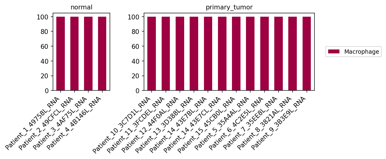
The plot_celltype_population tool was utilized to visualize the distribution of cells annotated as 'Macrophage' (from the celltype_minor category) across individual samples derived from both 'normal' and 'primary_tumor' breast tissue conditions. The objective of this analysis was to examine the population composition specifically within the macrophage cell type.
Visual Summary
The generated bar plot presents two distinct panels, segmenting the data by 'normal' and 'primary_tumor' conditions. Each panel displays multiple bars, with the x-axis representing individual patient samples (e.g., "Patient_1_49758L_RNA" for normal tissue, "Patient_10_3C7D1L_RNA" for primary tumor tissue), and the y-axis indicating a percentage from 0 to 100.
- Normal Samples: The panel for 'normal' condition shows four distinct samples. For each of these samples, a single burgundy-colored bar extends to the 100% mark on the y-axis. The legend explicitly identifies this color as representing "Macrophage" cells.
- Primary Tumor Samples: The panel for 'primary_tumor' condition displays ten distinct samples. Consistent with the normal samples, each primary tumor sample also features a single burgundy-colored bar reaching 100% on the y-axis, indicating the presence of "Macrophage" cells.
Biological Interpretation
- This plot serves as a confirmation that cells positively identified as 'Macrophage' are present in all interrogated normal and primary breast tumor samples within the dataset.
- The observation that all bars consistently reach 100% for 'Macrophage' is a direct consequence of the specific parameters used for this plot. The analysis was configured to targets': {'obs_col': 'celltype_minor', 'value': 'Macrophage'}, meaning the data was pre-filtered to include *only* cells classified as 'Macrophage' at the celltype_minor level. When a population plot is generated for a single, pre-selected cell type, that cell type will inherently constitute 100% of the displayed cells within its own category for each sample.
- Therefore, this visualization primarily confirms the successful annotation and presence of macrophage cells across the various samples and conditions. It does not, in its current presentation, offer insights into:
- The *relative abundance* or overall proportion of macrophages compared to all other cell types within each sample or across conditions.
- The *heterogeneity* or specific *subtypes* of macrophages (e.g., M1-like, M2-like, or other polarization states) within the normal or tumor microenvironments. To resolve macrophage heterogeneity, the celltype_subset column (which includes annotations such as "Macrophage (M1)", "Macrophage (M2A)", etc.) would need to be utilized in the analysis configuration.
Clinical or Translational Implications
- While macrophages are recognized as essential immune components in both healthy tissues and the tumor microenvironment (where they are often termed Tumor-Associated Macrophages or TAMs), this specific plot provides limited direct clinical or translational implications.
- To derive more actionable clinical insights, further analyses would typically be required to investigate:
- Whether there are statistically significant differences in the *overall infiltration* of macrophages between normal breast tissue and primary tumors.
- The *proportions and functional states* of different macrophage subtypes (e.g., pro-inflammatory M1 versus immunosuppressive M2) within the tumor context, as these can significantly influence disease progression, prognosis, and responsiveness to various therapeutic strategies in breast cancer [1, 2].
- The current plot acts as a foundational check, verifying the presence of macrophages in the samples, which is a prerequisite for such more detailed and clinically relevant investigations into their roles in breast cancer pathogenesis.
---
References:
[1] PubMed search for "macrophage tumor breast cancer M1 M2" (PubMed Search)
[2] PubMed search for "macrophage polarization cancer prognosis" (PubMed Search)
10. Ploidy Population Analysis in Tumor-Origin and Unassigned Cells of Breast Tissue
[Analysis Visualization Results]...
Analysis Overview
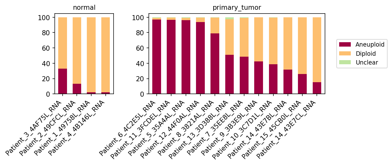
This analysis investigates the ploidy status (Aneuploid, Diploid, Unclear) of cells identified as "Epithelial cell" (the designated tumor-origin cell type for breast tissue) or "unassigned" cells within various patient samples from both normal breast tissue and primary breast tumors. The visualization provides a stacked bar plot, with each bar representing a single patient sample and showing the proportional distribution of ploidy states for the target cell populations within that sample. This helps to identify overall trends in chromosomal stability across disease conditions and patient samples.
Visual Summary
The bar plot clearly delineates the ploidy distribution in target cells (Epithelial and unassigned cells) between normal and primary tumor conditions.
- Normal Condition: In the normal breast tissue samples, the vast majority of target cells are classified as Diploid (light orange). A small proportion of Aneuploid cells (dark red) is observed in some normal samples (e.g., Patient_3_4AF75L_RNA, Patient_2_49CFCL_RNA), but they generally constitute less than ~35% of the population, and often much less or none. The 'Unclear' category is negligible.
- Primary Tumor Condition: In contrast, primary tumor samples exhibit a significantly higher proportion of Aneuploid cells. Many tumor samples (e.g., Patient_6_4C2E5L_RNA, Patient_11_3FCDEL_RNA, Patient_5_35A4AL_RNA, Patient_12_44F0AL_RNA) show near 100% aneuploidy in the target cell population. However, there is notable inter-sample variability within the primary tumor group. Some tumor samples (e.g., Patient_7_35EE8L_RNA, Patient_9_3B3E9L_RNA, Patient_10_3C7D1L_RNA, Patient_14_43E7BL_RNA, Patient_15_45CB0L_RNA, Patient_14_43E7CL_RNA) show a more mixed population, with Diploid cells making up a substantial fraction (ranging from ~20% to over ~80%). The 'Unclear' category remains very small in tumor samples as well.
Biological Interpretation
The observed shift in ploidy from predominantly diploid in normal samples to significantly aneuploid in primary tumor samples, particularly within the Epithelial cell population (tumor-origin cells), is a well-established hallmark of cancer.
- Aneuploidy as a Cancer Hallmark: Aneuploidy, the presence of an abnormal number of chromosomes, is a pervasive feature of most human cancers, including breast cancer. It often arises from chromosomal instability during cell division and can contribute to tumor initiation, progression, and heterogeneity by altering gene dosage and affecting cellular fitness [1].
- [1] PubMed search for "aneuploidy cancer" and "chromosomal instability cancer": https://pubmed.ncbi.nlm.nih.gov/?term=aneuploidy+cancer+chromosomal+instability
- Tumor-Origin Cells (Epithelial Cells): Given that epithelial cells are the origin of breast carcinomas, the high aneuploidy observed in these cells within primary tumors strongly suggests that these are indeed malignant epithelial cells undergoing oncogenic transformation and expansion. The presence of some aneuploidy in normal tissue might reflect age-related changes, early pre-malignant alterations, or technical artifacts, but its prevalence is drastically lower than in tumors.
- Sample Heterogeneity: The substantial variability in aneuploidy levels among different primary tumor samples is biologically significant. It could reflect:
- Tumor Purity: Samples with a higher proportion of diploid cells might have a greater infiltration of normal stromal or immune cells that are not aneuploid, or they might contain benign epithelial cells mixed with malignant ones.
- Tumor Evolution/Subtypes: Different breast cancer subtypes or stages of evolution may exhibit varying degrees of aneuploidy. Some tumors might be more chromosomally stable, while others are highly unstable.
- Intratumor Heterogeneity: A single tumor can contain subclones with different ploidy states, contributing to the overall mixed signal observed in bulk analysis or when pooling single-cell data from complex tumor microenvironments.
- "Unassigned" Cells: While the plot combines "Epithelial cell" and "unassigned", it's reasonable to infer that a significant portion of the aneuploid population in primary tumors originates from malignant epithelial cells. If some "unassigned" cells are also malignant cells that couldn't be definitively classified, they would contribute to this aneuploid signal. If "unassigned" cells largely represent non-malignant cells, their inclusion would dilute the aneuploid signal from the true tumor cells, making the observed high aneuploidy even more pronounced for the actual malignant population.
Clinical or Translational Implications
- Diagnostic and Prognostic Biomarker: Aneuploidy is a well-established prognostic marker in many cancers, including breast cancer. Higher degrees of aneuploidy or specific chromosomal aberrations can be associated with more aggressive disease, higher recurrence rates, or resistance to certain therapies.
- Therapeutic Targeting: Understanding the prevalence and nature of aneuploidy in a tumor can inform therapeutic strategies. For instance, cells with extensive aneuploidy might be more vulnerable to treatments that exploit chromosomal instability (e.g., agents affecting mitosis or DNA repair pathways). The variability observed suggests that a one-size-fits-all approach based solely on ploidy might not be effective for all breast cancer patients.
- Monitoring Tumor Evolution: Tracking ploidy changes over time could provide insights into tumor evolution and response to treatment. The presence of a significant diploid population within a primary tumor might suggest a less advanced stage or a subtype with lower chromosomal instability, warranting further investigation.
11. Primary Tumor vs. Normal Cell-Cell Interaction Patterns in Breast Tissue
[Analysis Visualization Results]...
Analysis Overview
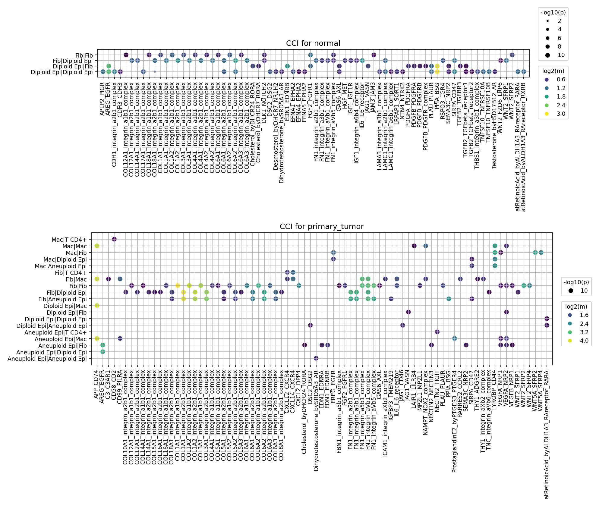
This analysis investigates cell-cell interaction (CCI) patterns in breast tissue, comparing 'normal' and 'primary_tumor' conditions. The plot_cci_dots tool was used to visualize significant ligand-receptor interactions between key cell types: Epithelial cells (distinguished by ploidy into Diploid and Aneuploid), Fibroblasts, Macrophages, and T cells (CD4+ and CD8+). Up to 80 significant interactions (p-value < 0.05, mean expression > 0.01) were selected for visualization per condition, revealing distinct cellular communication landscapes in healthy and cancerous breast microenvironments.
Visual Summary
Normal Condition:
- The interaction landscape in normal tissue is predominantly driven by Fibroblast-Fibroblast (Fib/Fib), Fibroblast-Diploid Epithelial (Fib/Diploid Epi), and Diploid Epithelial-Diploid Epithelial (Diploid Epi/Diploid Epi) cell pairs.
- Interactions are largely centered around extracellular matrix (ECM) components and their receptors, particularly various collagen-integrin complexes (e.g., COL1A1, COL1A2, COL3A1, COL4A1, COL5A1, COL6A1-3 interacting with integrin_a1b1, a2b1, a3b1, a11b1 complexes) and fibronectin (FN1)-integrin complexes. These interactions exhibit high significance and strong expression.
- Other notable interactions include laminin (LAMA3, LAMC1)-integrin complexes, PDGF signaling (PDGFA/PDGFRB, PDGFB/PDGFRB), TGFB2-TGFB Receptor complexes, and WNT2-SFRP4, suggesting roles in stromal maintenance and tissue homeostasis.
Primary Tumor Condition:
- The tumor microenvironment presents a more complex and diverse interaction network, involving Macrophages (Mac), Aneuploid Epithelial cells (Aneuploid Epi), and T cells (CD4+, CD8+) in addition to Fibroblasts and Diploid Epithelial cells.
- New cell-cell interaction pairs become prominent, such as Macrophage-Macrophage (Mac/Mac), Macrophage-Fibroblast (Mac/Fib), Macrophage-Epithelial (Mac/Diploid Epi, Mac/Aneuploid Epi), and interactions involving Aneuploid Epithelial cells with other tumor-associated cells.
- Key interactions that emerge or are significantly upregulated in the tumor include:
- MIF (Macrophage Migration Inhibitory Factor) interacting with CD74 and CXCR4: This axis is highly active across multiple Macrophage-involved cell pairs (Mac/Mac, Mac/Fib, Mac/Diploid Epi, Mac/Aneuploid Epi).
- SPP1 (Osteopontin) interacting with CD44 and integrin_a4b1_complex: Prominent in Macrophage-Epithelial and Macrophage-Macrophage interactions.
- TNC (Tenascin C) interacting with integrin_a9b1_complex and integrin_avb6_complex: Strong between Fibroblasts and Epithelial cells (both Diploid and Aneuploid) and Fibroblast-Fibroblast pairs.
- IL6-IL6R signaling: Highly active in Macrophage-Macrophage interactions.
- PGE2 (Prostaglandin E2)-PTGER4: Strong in Macrophage-Macrophage interactions.
- APP (Amyloid Beta Precursor Protein)-CD74/CD99/PIRB: Strong in Macrophage-Macrophage interactions.
- The continued presence of collagen-integrin and fibronectin-integrin complexes suggests ongoing ECM remodeling, but likely in a pathological context.
- PDGF, TGFB2, and WNT pathways (WNT2/SFRP4, WNT7A/SFRP4, WNT5A/FZD) remain active or are dysregulated in the tumor.
- ICAM1-LFA-1 complex interactions are observed, particularly between Fibroblasts and T cells.
Biological Interpretation
The comparison highlights a dramatic shift in cellular communication from a homeostatic state in normal breast tissue to a pro-tumorigenic microenvironment.
- ECM Remodeling and Stromal Support: In normal tissue, strong collagen and fibronectin interactions underscore the structural integrity and normal cell adhesion mediated by fibroblasts and epithelial cells. In the tumor, while these persist, the emergence of Tenascin C (TNC) interactions, particularly involving fibroblasts, signifies pathological extracellular matrix (ECM) remodeling, a hallmark of cancer-associated fibroblasts (CAFs) that support tumor growth and invasion. GeneCards - TNC
- Inflammation and Immunosuppression driven by Macrophages: The most striking difference in the tumor microenvironment is the widespread and robust engagement of macrophages.
- The MIF-CD74/CXCR4 axis is highly active. Macrophage Migration Inhibitory Factor (MIF) is a crucial cytokine involved in inflammation, immune evasion, and tumor progression, often secreted by tumor cells and macrophages themselves, fostering a pro-inflammatory and immunosuppressive milieu. PubMed search: MIF CD74 cancer
- SPP1 (Osteopontin) signaling via CD44 and integrins is significantly upregulated. SPP1, often produced by tumor-associated macrophages (TAMs) and tumor cells, promotes angiogenesis, metastasis, and suppresses anti-tumor immunity. GeneCards - SPP1
- The strong IL6-IL6R and PGE2-PTGER4 interactions also point to macrophage-driven inflammation that fuels tumor proliferation, survival, and immune evasion. PubMed search: IL6 PGE2 cancer microenvironment
- Interactions involving APP-CD74 suggest additional complex roles for macrophages in regulating the TME.
- Malignant Epithelial Cell Interactions: The distinct emergence of Aneuploid Epithelial cells interacting with other cell types (fibroblasts, macrophages, and other aneuploid epithelial cells) reflects the neoplastic transformation. These malignant cells likely engage in unique communication pathways that promote their survival, proliferation, and interaction with the stromal and immune components of the tumor.
Dysregulated Growth Factor and Developmental Signaling:
- PDGF signaling continues to be active, indicating ongoing fibroblast proliferation and stromal remodeling, critical for supporting tumor growth.
- TGFB2-TGFB Receptor complex interactions highlight the role of TGF-β in the TME, which can promote fibrosis, suppress anti-tumor immunity, and drive epithelial-mesenchymal transition (EMT). PubMed search: TGFB cancer TME
- The various WNT ligand-receptor interactions (WNT2, WNT7A, WNT5A) point to reactivation of developmental pathways that often contribute to cancer cell proliferation, stemness, and resistance to therapy.
- Immune Cell Adhesion: The ICAM1-LFA-1 interactions observed between fibroblasts and T cells indicate crucial adhesion events, important for immune cell trafficking and potential immune synapse formation within the TME, which can be either anti-tumorigenic or pro-tumorigenic depending on the context.
Clinical or Translational Implications
The identified specific ligand-receptor interactions provide critical insights for potential therapeutic interventions and biomarker development in breast cancer:
Therapeutic Target Prioritization:
- The MIF-CD74/CXCR4 axis and SPP1-CD44/integrin signaling emerge as high-priority targets. Given their central role in macrophage-driven inflammation, immunosuppression, angiogenesis, and metastasis within the tumor microenvironment, targeting these pathways (e.g., with small molecule inhibitors or blocking antibodies) could disrupt multiple pro-tumorigenic processes.
- Modulating Tenascin C (TNC) interactions could be a strategy to normalize the pathological ECM, thereby reducing tumor growth, invasion, and improving drug delivery.
- Interfering with IL6-IL6R, PGE2-PTGER4, or dysregulated WNT signaling could suppress inflammation and inhibit cancer cell proliferation and survival.
- Biomarker Discovery: The distinct CCI profiles observed between normal and tumor tissue, especially involving aneuploid epithelial cells and macrophages, could lead to the discovery of novel prognostic or predictive biomarkers. For instance, high expression or activity of MIF, SPP1, or TNC interactions could indicate more aggressive disease or predict response to specific therapies.
- Experimental Validation and Combination Strategies: The identified interactions provide a strong basis for further experimental validation.
- *In vitro* studies could involve functional assays (e.g., migration, invasion, immune cell activation) using cell lines or patient-derived organoids, coupled with gene knockdown/knockout or receptor blocking experiments.
- *In vivo* studies using patient-derived xenograft (PDX) models or syngeneic mouse models could assess the therapeutic efficacy of targeting these pathways, potentially in combination with existing therapies (e.g., chemotherapy, immunotherapy).
- Understanding the interplay between T cells and other TME components (e.g., ICAM1-LFA-1) could inform strategies for optimizing adoptive cell therapies or immune checkpoint blockade.
12. Normal vs. Tumor Microenvironment: Comparative Analysis of Cell-Cell Interactions in Breast Tissue
[Analysis Visualization Results]...
Analysis Overview
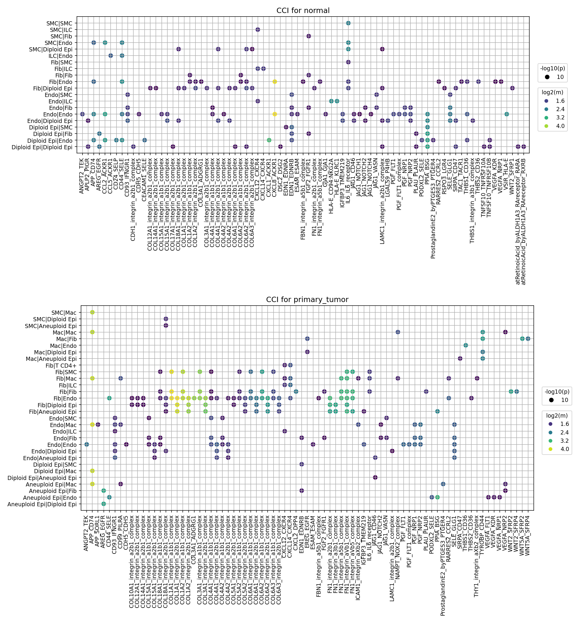
This analysis investigates cell-cell interactions (CCIs) in breast tissue, comparing normal and primary tumor conditions using single-cell RNA sequencing data. The plot_cci_dots tool, based on CellPhoneDB, was utilized to identify and visualize significant ligand-receptor interactions between different cell types. The plots display the top 80 interacting gene pairs for each condition, with dot size representing the statistical significance (-log10(p-value)) and dot color indicating the interaction strength (log2(mean expression)). The analysis specifically highlights interactions involving normal (Diploid) and malignant (Aneuploid) epithelial cells, along with various stromal and immune cell types.
Visual Summary
- CCI for Normal Breast Tissue:
- Key Cell Pairs: Interactions are predominantly observed between Fibroblasts (Fib) and Epithelial cells (Diploid Epi), Endothelial cells (Endo) and Epithelial cells (Diploid Epi), and various stromal components like Smooth Muscle Cells (SMC) and Endothelial cells.
- Dominant Ligand-Receptor Systems: A prominent feature is the extensive array of collagen-integrin interactions (e.g., COL1A1/COL1A2/COL1A3-integrin_a1b1/a2b1/a3b1_complexes). These interactions, involving various collagen types and integrin subunits, are widespread across multiple cell pairs, reflecting the critical role of extracellular matrix (ECM) components in maintaining tissue structure and cellular communication in healthy tissue.
- Other Noted Interactions: Other interactions include EGF-EGFR, CXCL12-CXCR4, FGF-FGFR1, and ICAM1-ITGAL/ITGB2, suggesting basal levels of growth factor signaling, chemokine signaling, and cell adhesion.
- Ploidy: Epithelial cells in the normal condition are primarily identified as Diploid Epi, consistent with healthy tissue.
- CCI for Primary Tumor Breast Tissue:
- Emergence of Aneuploid Epithelial Cells: A critical difference is the presence of Aneuploid Epi cells, which are malignant epithelial cells. These cells engage in numerous interactions with Fibroblasts, Macrophages, and Endothelial cells, highlighting the remodeled tumor microenvironment.
- Shift in Key Cell Pairs: Macrophage (Mac) interactions become highly prominent, especially with Epithelial cells (Mac/Diploid Epi, Mac/Aneuploid Epi) and Fibroblasts (Mac/Fib). Interactions involving T cells (Fib/T CD4+) also appear, indicating immune cell infiltration.
Altered Ligand-Receptor Landscape:
- Sustained ECM Interactions: Collagen-integrin and Fibronectin-integrin (FN1_integrin_a2b1/a5b1_complexes) interactions remain highly prevalent, often with high mean expression, suggesting ongoing and possibly exaggerated ECM remodeling within the tumor.
- Pro-angiogenic Signaling: VEGFA-FLT1 and VEGFA-KDR interactions are clearly visible, particularly involving Endothelial cells, indicative of active angiogenesis.
- Tumor Progression-related Signaling: Interactions involving SPP1 (secreted phosphoprotein 1, also known as Osteopontin) with integrin complexes are notable, often with high mean expression. WNT5A-FZD (FZD2, FZD5, FZD8) interactions, representing non-canonical Wnt signaling, also emerge.
- Immune/Inflammatory Mediators: CXCL12-CXCR4 continues to be present, and interactions such as ANG2-TEK (Tie2 signaling) are also observed.
- Increased Diversity and Strength: The primary tumor plot generally shows a more diverse set of interacting cell pairs and ligand-receptor complexes, often with high mean expression values (more yellow/green dots), suggesting a more active and complex interactive network.
Biological Interpretation
The comparison between normal and primary tumor conditions reveals significant shifts in cell-cell communication, reflecting the hallmarks of cancer development and progression.
- ECM Remodeling and Fibrosis: In both conditions, collagen-integrin interactions are fundamental. However, their pervasive presence and high expression in the tumor context, alongside fibronectin-integrin and SPP1-integrin interactions, indicate extensive and likely dysregulated ECM remodeling. This stiffened and altered ECM (e.g., via SPP1 signaling) can promote tumor cell proliferation, survival, migration, and metastasis, while also influencing immune cell behavior within the tumor microenvironment [1, 2].
- Angiogenesis: The strong presence of VEGFA-FLT1/KDR (VEGFR1/VEGFR2) interactions in the primary tumor is a classic signature of tumor angiogenesis. Malignant epithelial cells and stromal cells secrete VEGFA, stimulating endothelial cell proliferation and migration to form new blood vessels, which are crucial for supplying nutrients and oxygen to the rapidly growing tumor [3].
- Immune Microenvironment Alterations: The dramatic increase in macrophage-epithelial cell interactions in the tumor suggests significant immune cell infiltration and cross-talk. Macrophages, particularly tumor-associated macrophages (TAMs), are often polarized towards an M2-like phenotype in tumors and can promote tumor growth, angiogenesis, and immunosuppression [4]. The presence of Fibroblast-T cell (CD4+) interactions also indicates active immune surveillance or suppression within the tumor.
- Growth Factor and Chemokine Signaling: Upregulated EGF-EGFR and FGF-FGFR1 signaling can drive uncontrolled proliferation and survival of tumor cells. The CXCL12-CXCR4 axis is well-known for its roles in guiding cell migration (e.g., metastasis) and promoting tumor cell survival [5].
- Non-canonical Wnt Signaling: The emergence of WNT5A-FZD interactions in the tumor suggests activation of non-canonical Wnt pathways. WNT5A signaling is often implicated in promoting tumor cell migration, invasion, and metastasis in various cancers, including breast cancer [6].
- Role of Aneuploid Epithelial Cells: The specific interactions involving Aneuploid Epi cells underscore their active role in shaping the tumor microenvironment by directly communicating with stromal and immune cells. These interactions are critical for their survival, proliferation, and malignant progression.
Clinical or Translational Implications
The identified cell-cell interactions represent a rich source of potential therapeutic targets and biomarkers for breast cancer.
- Targeting Angiogenesis: The strong VEGFA-VEGFR signaling confirms angiogenesis as a crucial hallmark in breast cancer. Anti-angiogenic therapies targeting VEGF or its receptors (e.g., bevacizumab for VEGFA, sunitinib/pazopanib for VEGFRs) are established treatments in various cancers and could be further optimized or explored in specific breast cancer contexts [7].
- Modulating ECM Remodeling: Ligand-receptor pairs involving collagens, fibronectin, and particularly SPP1 with integrins, present opportunities to disrupt tumor-promoting ECM interactions. SPP1, often highly expressed in aggressive cancers, is a promising target for inhibiting tumor progression, metastasis, and immune evasion [2]. Inhibitors against specific integrin subunits (e.g., αvβ3, α5β1) involved in these interactions could impede tumor growth and metastasis [8].
- Reprogramming the Immune Microenvironment: Targeting macrophage-epithelial interactions, or specific pathways like CXCL12-CXCR4, could reprogram TAMs from a pro-tumorigenic to an anti-tumorigenic state, or block immune cell recruitment that supports tumor growth.
- Inhibiting Growth and Survival Pathways: The continued presence of EGF-EGFR and FGF-FGFR1 interactions highlights the potential for existing targeted therapies (e.g., EGFR inhibitors like gefitinib or erlotinib) to be effective, or for novel inhibitors against other growth factor receptors to be developed.
- Wnt Pathway Modulation: Targeting WNT5A-FZD interactions could disrupt non-canonical Wnt signaling, which is implicated in metastasis and resistance to therapy. Wnt pathway inhibitors are an active area of drug development in oncology [9].
- Biomarker Discovery: The specific ligand-receptor pairs highly expressed and active in the primary tumor, particularly those involving Aneuploid Epi cells, could serve as prognostic biomarkers for disease aggressiveness or predictive markers for response to specific targeted therapies.
---
References:
- Collagen-integrin interactions in cancer: For a general overview of integrins in cancer:
PubMed Search: "integrin cancer" review
- SPP1 (Osteopontin) in cancer: For its role in ECM remodeling, immunity, and cancer progression:
PubMed Search: "SPP1 cancer" review
- VEGF-VEGFR signaling in angiogenesis: For details on angiogenesis in cancer:
PubMed Search: "VEGF angiogenesis cancer" review
- Tumor-associated macrophages: For their role in the tumor microenvironment:
PubMed Search: "tumor associated macrophages" review
- CXCL12-CXCR4 axis in cancer: For its role in cell migration and metastasis:
- WNT5A in cancer: For its role in non-canonical Wnt signaling and metastasis:
PubMed Search: "WNT5A cancer" review
- Anti-angiogenic therapies in cancer: For clinical implications of VEGF targeting:
PubMed Search: "anti-VEGF therapy cancer" review
- Integrin inhibitors in cancer therapy: For therapeutic potential of targeting integrins:
PubMed Search: "integrin inhibitor cancer therapy" review
- Wnt pathway inhibitors in cancer: For therapeutic approaches targeting Wnt signaling:
PubMed Search: "Wnt pathway inhibitor cancer" review
13. Immune Checkpoint and Cell Cycle Related Cell-Cell Interactions in Breast Tissue
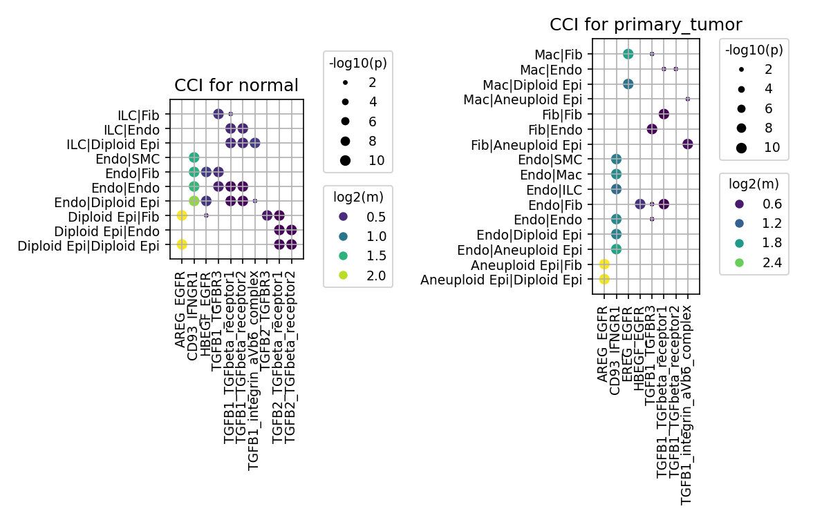
[Analysis Visualization Results]...
Analysis Overview
This analysis investigates cell-cell interactions (CCI) within breast tissue, specifically focusing on a predefined set of genes related to immune checkpoints and cell cycle pathways. The plot_cci_dots tool was used to visualize significant ligand-receptor interactions between various cell types in both normal and primary tumor conditions. While the initial query included a broad list of immune checkpoint and cell cycle genes, the resulting plots highlight specific interactions involving EGFR signaling (AREG-EGFR, HBEGF-EGFR, EREG-EGFR), IFN-gamma signaling (CD93-IFNGR1), and TGF-beta signaling (TGFB1/2-TGFBR1/2/3, TGFB1-integrin_avb6_complex) that met the significance and mean expression cutoffs. The analysis distinguishes between Diploid Epithelial cells and Aneuploid Epithelial cells, reflecting the ploidy inference for the tumor origin cell type.
Visual Summary
CCI for Normal Condition
- Dominant Interactions: In normal breast tissue, TGF-beta signaling pathways (TGFB1/2 interacting with various TGF-beta receptors) show widespread and highly significant interactions (large, dark purple/green/yellow dots), particularly between ILCs, Endothelial cells, Fibroblasts, and Diploid Epithelial cells.
- EGFR Signaling: AREG-EGFR and HBEGF-EGFR interactions are present, notably involving Diploid Epithelial cells with Endothelial cells and Fibroblasts, suggesting paracrine/autocrine growth factor support.
- Immune Signaling: CD93-IFNGR1 interactions are observed, primarily between ILCs and Fibroblasts/Endothelial cells, indicating immune-stromal cross-talk.
- Key Cell Types: ILCs, Fibroblasts, Endothelial cells, and Diploid Epithelial cells are the primary participants in these significant interactions.
CCI for Primary Tumor Condition
- Enhanced TGF-beta Signaling: TGF-beta signaling remains a prominent interaction axis, often with increased significance (larger dots) and higher mean expression (more yellow/light green colors) compared to normal. Key interactions involve Fibroblasts with Aneuploid Epithelial cells and Endothelial cells, and also Aneuploid Epithelial cells interacting with Diploid Epithelial cells. The presence of TGFB1-integrin_avb6_complex highlights a potentially distinct mechanism of TGF-beta activation or presentation in the tumor microenvironment.
- Activated EGFR Signaling: EGFR ligand-receptor pairs (AREG-EGFR, HBEGF-EGFR, EREG-EGFR) show strong interactions, particularly involving Aneuploid Epithelial cells with Fibroblasts and Diploid Epithelial cells, and also within Endothelial cells. EREG-EGFR appears to be more prominent in the tumor context, notably in Aneuploid Epi|Diploid Epi and Aneuploid Epi|Fib interactions.
- Macrophages in Tumor Microenvironment: Macrophages emerge as significant interactors in the tumor, showing connections with Fibroblasts, Diploid Epithelial cells, and Endothelial cells, including interactions involving CD93-IFNGR1.
- Aneuploid Epithelial Cells as Key Players: Aneuploid Epithelial cells (representing tumor cells) are highly active in signaling, engaging extensively with Fibroblasts and Diploid Epithelial cells through both EGFR and TGF-beta pathways.
Key Differences Between Normal and Tumor
- Shift in Cell Type Participation: Macrophages become more active in CCI in the tumor. Crucially, Aneuploid Epithelial cells show extensive interactions, highlighting their role in shaping the tumor microenvironment.
- Intensification of Growth Factor and Immunomodulatory Signaling: Both EGFR and TGF-beta signaling pathways appear to be intensified in the primary tumor, often with higher mean expression and significance, especially involving tumor-associated fibroblasts and aneuploid epithelial cells.
- Emergence of Specific Complexes: The TGFB1-integrin_avb6_complex interaction specifically observed in the tumor context suggests a potentially unique mechanism of TGF-beta signaling activation or receptor binding relevant to cancer progression.
- Absence of Canonical Immune Checkpoint Ligand-Receptor Pairs: Despite the query for immune checkpoint genes, the generated plots do not display interactions such as PD1-PDL1 (CD274) or CTLA4-CD80/CD86. This indicates that these specific pairs might not have met the significance and mean expression cutoffs within this analysis, or were not among the top interactions selected for display by the tool.
Biological Interpretation
The observed cell-cell interactions underscore critical aspects of breast cancer biology and its microenvironment.
- EGFR Signaling in Tumor Progression: The upregulation and broadened engagement of EGFR ligands (AREG, HBEGF, EREG) with EGFR, particularly involving Aneuploid Epithelial cells in the tumor, highlight the autocrine and paracrine loops driving tumor cell proliferation, survival, and migration. EGFR is a well-established oncogenic driver in various cancers, and its activation in tumor cells and stromal components suggests its central role in tumor growth and metastasis. The prominence of EREG-EGFR in the tumor context could indicate a specific mechanism of EGFR activation in breast cancer progression 1.
- TGF-beta Signaling as a Double-Edged Sword: TGF-beta signaling is intensely active in both normal and tumor conditions. While crucial for tissue homeostasis in normal tissue, its dysregulation in cancer can promote epithelial-to-mesenchymal transition (EMT), immune suppression, angiogenesis, and extracellular matrix remodeling, contributing to tumor progression. The strong interactions between Aneuploid Epithelial cells, Fibroblasts, and Endothelial cells via TGF-beta suggest its critical role in orchestrating the tumor microenvironment. The appearance of the TGFB1-integrin_avb6_complex could indicate context-specific activation of TGF-beta, as integrin αvβ6 can activate latent TGF-β, driving pro-tumorigenic effects 2.
Role of Tumor Microenvironment (TME) Cells
- Fibroblasts: Their extensive interactions, especially with Aneuploid Epithelial cells and Endothelial cells, through both EGFR and TGF-beta pathways, confirm their role as critical components of the TME, actively supporting tumor growth and remodeling.
- Macrophages: The increased involvement of macrophages in tumor CCI, including CD93-IFNGR1 interactions, points to their integration into the TME. Tumor-associated macrophages (TAMs) often adopt an M2-like phenotype that promotes tumor growth, angiogenesis, and immune suppression 3.
- Endothelial Cells: Their persistent and often intensified interactions in the tumor suggest their crucial role in angiogenesis, supporting the nutrient supply for the growing tumor.
- Ploidy Status: The distinction between Diploid and Aneuploid Epithelial cells in the tumor context is highly informative. The Aneuploid Epithelial cells (likely the malignant clones) exhibit distinct and often stronger pro-tumorigenic signaling (e.g., EREG-EGFR, TGFB-integrin_avb6_complex) compared to their diploid counterparts, highlighting their active role in establishing and maintaining the tumor phenotype.
Clinical or Translational Implications
The findings from this CCI analysis provide several potential clinical and translational avenues:
- Therapeutic Targeting of EGFR Pathway: The consistent and intensified activation of EGFR signaling in the primary tumor, particularly involving Aneuploid Epithelial cells, reinforces EGFR as a validated therapeutic target in breast cancer. Given the specific activity of EREG-EGFR, therapeutic strategies specifically inhibiting EREG or its downstream signaling could be explored. Existing EGFR inhibitors (e.g., gefitinib, erlotinib) might be effective in subsets of breast cancer characterized by high EREG-EGFR activity 4.
- TGF-beta Pathway as a Therapeutic Target: The widespread and often increased TGF-beta signaling in the tumor microenvironment suggests its potential as a therapeutic target to inhibit tumor progression, metastasis, and immune evasion. Developing or repurposing drugs that block TGF-beta signaling (e.g., small molecule inhibitors, antibodies) could be beneficial, especially considering the interactions involving tumor cells and stromal components like fibroblasts 5. The specific targeting of integrin αvβ6, which activates TGF-β, could offer a more selective therapeutic approach to mitigate pro-tumorigenic TGF-β effects.
- Targeting Tumor Microenvironment: The active participation of Fibroblasts and Macrophages in key signaling pathways (EGFR, TGF-beta) highlights the importance of targeting the tumor microenvironment. Combining therapies that target tumor cells with those that modulate stromal cells could yield more effective anti-cancer responses.
- Biomarker Discovery: The distinct CCI patterns, particularly those involving Aneuploid Epithelial cells and specific ligand-receptor pairs (e.g., EREG-EGFR, TGFB1-integrin_avb6_complex), could serve as prognostic or predictive biomarkers for patient stratification and response to targeted therapies.
- Further Experimental Validation: The identified critical interactions warrant further experimental validation using in vitro co-culture models, organoids, or in vivo animal models to confirm their functional roles in breast cancer progression and therapeutic response. This could involve blocking specific ligands or receptors to assess their impact on tumor growth, invasion, and immune modulation.
14. Condition-Specific Cell-Cell Interaction Patterns in Breast Tissue
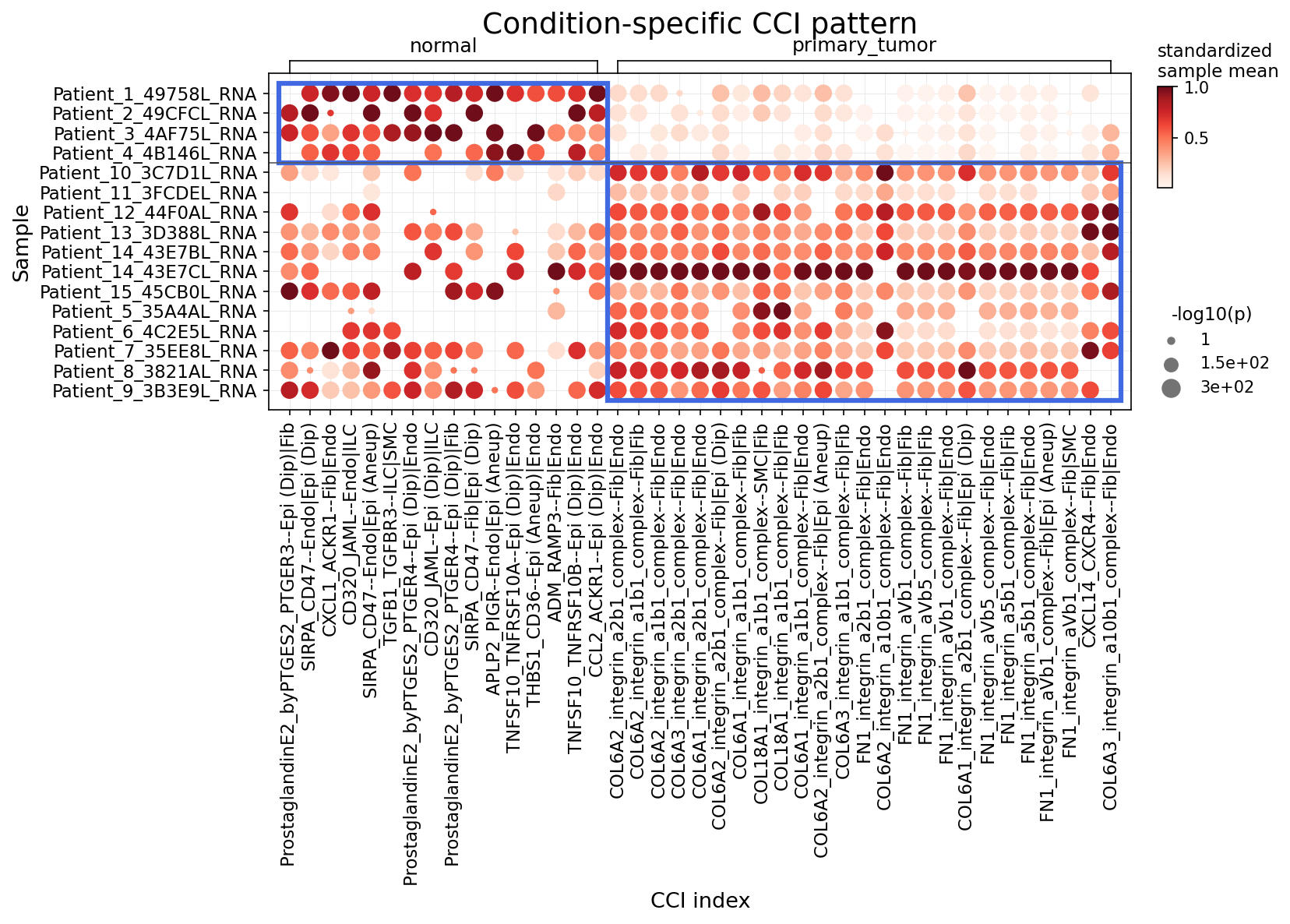
[Analysis Visualization Results]...
Analysis Overview
This analysis investigates statistically significant differences in cell-cell interactions (CCIs) between normal breast tissue and primary_tumor conditions, focusing on major immune and stromal cells, as well as epithelial cells. The results are visualized as a dot plot, where the size of each dot represents the statistical significance (-log10(p-value)) of the interaction, and the color intensity indicates the standardized mean interaction strength across samples. The plot highlights the top 25 most significant CCI pairs in each condition group, allowing for a comparative assessment of the tumor microenvironment versus normal tissue.
Visual Summary
The dot plot, "Condition-specific CCI pattern," effectively illustrates distinct CCI landscapes in normal versus primary tumor breast tissue.
- Condition-Specific Clustering: The plot clearly separates into two major panels: normal and primary_tumor. The CCI pairs enriched in normal samples are predominantly observed in the left panel, while a largely distinct set of interactions is highly active in the primary tumor samples, shown in the right panel.
Normal Tissue Interactions
- A prominent cluster of strong and significant interactions (dark red, large dots) is visible in the normal condition, particularly for the first few patient samples (Patient_1_49758L_RNA to Patient_10_3C7D1L_RNA).
- Key interaction pairs in normal tissue involve various cell types including Epithelial (both Diploid (Dip) and Aneuploid (Aneup)), Fibroblast, Endothelial, and ILCs.
- Notable interactions include ProstaglandinE2_byPTGES2-PTGER3 (Epithelial(Dip)-Fibroblast), CXCL1-ACKR1 (Fibroblast-Endothelial), CD47-Endo (Endothelial-Epithelial(Dip)), SIRPA_CD47 (Endothelial-Epithelial(Aneuploid)), TGFB1_TGFBR3 (ILC-Smooth muscle cell), TNFSF10_TNFRSF10A (Epithelial(Dip)-Endothelial), and CCL2_ACKR1 (Epithelial(Dip)-Endothelial).
Primary Tumor Interactions
- The primary_tumor condition exhibits a distinct set of highly active and significant CCIs. These interactions are broadly present across most tumor samples, indicating common features of the tumor microenvironment.
- A striking feature is the dominance of integrin interactions with collagen (COL6A, COL18A) and fibronectin (FN1). These interactions involve Fibroblasts, Endothelial cells, Epithelial cells (both Diploid and Aneuploid), and Smooth muscle cells.
- Specific examples include COL6A2_integrin_a2b1_complex (Fibroblast-Fibroblast, Fibroblast-Endothelial), COL6A1_integrin_a1b1_complex (Fibroblast-Endothelial), and FN1_integrin_aVb5_complex (Fibroblast-Fibroblast).
- The CXCL14-CXCR4 axis between Fibroblasts and Endothelial cells is also prominent in tumor samples.
- Ploidy Status: Some interactions specify the ploidy of epithelial cells (e.g., Epi(Dip), Epi(Aneup)), suggesting potential differences in how diploid vs. aneuploid epithelial cells engage in CCIs within the microenvironment. For instance, CD47-Endo interacts with Epithelial(Diploid), while SIRPA_CD47 interacts with Epithelial(Aneuploid).
Biological Interpretation
The observed condition-specific CCI patterns provide critical insights into the dynamic interplay within the breast tumor microenvironment compared to normal tissue homeostasis.
- Normal Tissue Homeostasis and Immune Surveillance: The normal breast tissue displays a diverse array of interactions.
- The presence of ProstaglandinE2-PTGER3 signaling, often involved in inflammation and immune regulation, suggests active immune responses or tissue remodeling processes, even in normal tissue [1]. Prostaglandins can modulate immune cell function.
- CXCL1-ACKR1 and CCL2-ACKR1 interactions point to chemokine signaling, crucial for immune cell trafficking and modulating the local immune landscape [2]. ACKR1 (DARC) is a chemokine receptor known to regulate chemokine availability.
- CD47-SIRPA interactions are crucial "don't eat me" signals that prevent phagocytosis of healthy cells by macrophages [3]. This interaction often involves Epithelial cells (both Diploid and Aneuploid) and Endothelial cells, suggesting a mechanism to maintain tissue integrity and immune evasion, which can be co-opted by cancer cells.
- TGFB1-TGFBR3 signaling can play roles in immune suppression and fibrogenesis. Its involvement with ILCs and smooth muscle cells suggests a regulatory role in the normal tissue microenvironment.
- TNFSF10-TNFRSF10A (TRAIL-TRAILR1) interactions can induce apoptosis and might represent part of immune surveillance mechanisms or cell turnover processes [4].
- Tumor Microenvironment Remodeling and Progression: The striking shift in CCIs in primary tumors highlights a profound reorganization of the extracellular matrix (ECM) and cell adhesion landscape, driven largely by cancer-associated fibroblasts (CAFs) and endothelial cells.
- ECM Remodeling and Stiffness: The overwhelming abundance of integrin interactions with collagen (COL6A, COL18A) and fibronectin (FN1) is a hallmark of the tumor microenvironment [5].
- Integrins are cell surface receptors that mediate cell-ECM and cell-cell adhesion, critical for cell migration, proliferation, and survival.
- Collagen VI (COL6A) and Fibronectin (FN1) are major components of the ECM. Increased deposition and cross-linking of these proteins lead to ECM stiffening, which promotes tumor progression, invasion, and metastasis by providing physical cues and signaling platforms for tumor cells and stromal cells [6, 7].
- Interactions like COL6A2_integrin_a2b1_complex (Fibroblast-Fibroblast, Fibroblast-Endothelial) suggest robust fibroblast activation and their crucial role in depositing and remodeling the ECM, forming a desmoplastic stroma.
- Angiogenesis and Metastasis: The CXCL14-CXCR4 axis between Fibroblasts and Endothelial cells is known to promote angiogenesis, tumor growth, and metastasis in various cancers, including breast cancer [8]. This axis facilitates communication between stromal and endothelial cells to support tumor vascularization.
- Ploidy-specific interactions: The distinct CD47 interactions based on epithelial cell ploidy suggest that aneuploid epithelial cells might engage in different immune evasion strategies or interact uniquely with their microenvironment compared to diploid epithelial cells.
Clinical or Translational Implications
The identified condition-specific CCI patterns have significant clinical and translational implications for breast cancer:
- Biomarker Discovery: The distinct sets of CCIs active in normal versus tumor tissue could serve as potential diagnostic or prognostic biomarkers. For instance, a high prevalence of specific integrin-ECM interactions (e.g., COL6A-integrin, FN1-integrin) could indicate tumor presence or aggressive behavior.
- Therapeutic Targets: The dominant integrin-ECM interactions and the CXCL14-CXCR4 axis in primary tumors represent promising therapeutic targets.
- Integrin Inhibition: Inhibitors targeting specific integrin subunits (e.g., α2β1, α1β1, αVβ5) could disrupt tumor cell adhesion, migration, and invasion, as well as normalize the tumor microenvironment [9].
- ECM-targeting therapies: Strategies to reduce ECM stiffness or degrade specific ECM components like Collagen VI or Fibronectin could impede tumor growth and improve drug delivery.
- CXCR4 Antagonists: Blocking the CXCL14-CXCR4 axis could inhibit tumor angiogenesis and metastatic spread, potentially synergizing with existing therapies [10].
- Understanding Treatment Resistance: Altered CCIs might also contribute to therapeutic resistance. For example, enhanced ECM stiffness can impair drug penetration or activate survival pathways in tumor cells.
- Immune Modulation: The differences in immune-related CCIs (e.g., CD47-SIRPA, Prostaglandin E2, TNFSF10) between conditions offer insights into immune evasion mechanisms in the tumor and potential targets for immunotherapies.
---
References:
[1] Prostaglandin E2 in cancer: A PubMed search for "prostaglandin E2 cancer immune" can provide context. E.g., PubMed Search
[2] Chemokine receptors ACKR1 and immune cell trafficking: A PubMed search for "ACKR1 chemokine immune trafficking". E.g., PubMed Search
[3] CD47-SIRPA "don't eat me" signal: GeneCards entry for CD47 or SIRPA provides a good overview. E.g., GeneCards
[4] TRAIL/TNFSF10 in cancer: A PubMed search for "TNFSF10 TRAIL apoptosis cancer". E.g., PubMed Search
[5] Integrins in cancer: GeneCards entry for Integrin. E.g., GeneCards or GeneCards
[6] Collagen VI in tumor microenvironment: A PubMed search for "collagen VI cancer microenvironment". E.g., PubMed Search
[7] Fibronectin in tumor microenvironment: A PubMed search for "fibronectin cancer microenvironment". E.g., PubMed Search
[8] CXCL14-CXCR4 axis in cancer: A PubMed search for "CXCL14 CXCR4 cancer angiogenesis". E.g., PubMed Search
[9] Integrin inhibitors in cancer therapy: A PubMed search for "integrin inhibitors cancer therapy". E.g., PubMed Search
[10] CXCR4 antagonists in cancer therapy: A PubMed search for "CXCR4 antagonists cancer therapy". E.g., PubMed Search
15. 유방암 상피세포의 조건별 표면 마커 분석
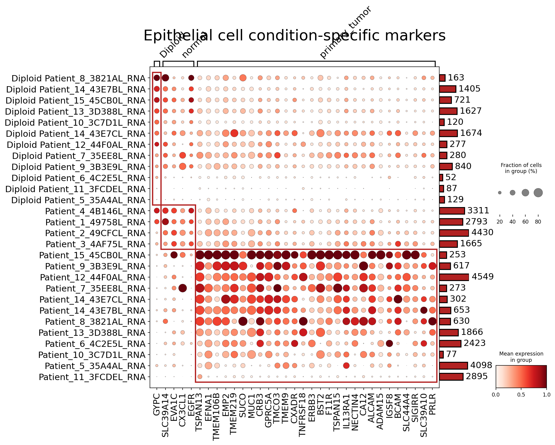
[Analysis Visualization Results]...
Analysis Overview
본 분석은 단일 세포 RNA 시퀀싱 데이터를 활용하여 유방 조직 내 상피세포(tumor-origin cells)에서 정상 조건과 원발성 종양(primary_tumor) 조건 간의 차등 발현되는 표면 마커를 식별하고 시각화합니다. 특히, 핵형(ploidy) 상태(이배체(Diploid) 또는 이수체(Aneuploid))에 따른 종양 샘플의 구분과 마커 발현 패턴을 확인합니다. 'plot_markers_and_expression_dot' 도구를 사용하여 조건별 최대 50개의 표면 마커가 선택되었으며, 이들의 발현 수준과 발현 세포 비율이 점도표로 표시되었습니다.
Visual Summary
점도표는 상피세포에서 정상 및 종양 조건에 따라 유의하게 차등 발현되는 표면 마커 유전자들의 평균 발현량(점의 색깔)과 발현 세포 비율(점의 크기)을 보여줍니다.
- 세포 집단 구분: Y축은 샘플들을 핵형(ploidy) 및 조건(normal/primary_tumor)에 따라 그룹화합니다.
- 상단 그룹: Diploid normal 샘플들로, 이배체 정상 상피세포를 나타냅니다.
- 중간 그룹: Diploid primary_tumor 샘플들로, 이배체 원발성 종양 상피세포를 나타냅니다.
- 하단 그룹: Patient_X_RNA로 명명된 샘플들로, 맥락상 이수체(Aneuploid) 원발성 종양 상피세포를 나타내는 것으로 추정됩니다.
정상 특이 마커:
- GYPC, SLC39A14, EVA1C, CX3CL1 등은 주로 Diploid normal 상피세포 그룹에서 높은 발현 수준과 넓은 발현 세포 비율을 보이며, 종양 샘플에서는 발현이 낮거나 거의 없습니다. 이는 이들이 정상 상피세포의 특징적인 표면 마커임을 시사합니다.
종양 특이 마커:
- 대부분의 유전자, 특히 EGFR, TSPAN13, EFNA1, TMEM106B, EMP2, MUC1, GPRC5A, TMEM9, CXADR, TNFRSF18, ERBB3, BST2, F11R, TSPAN15, IL13RA1, NECTIN4, CA12, ALCAM, ADAM15, IGSF8, BCAM, SLC4A4, SIGIRR, PRLR 등은 Diploid primary_tumor 및 Aneuploid primary_tumor 그룹에서 전반적으로 높은 발현을 보입니다.
- 특히 MUC1, GPRC5A, TMEM9, CXADR, TNFRSF18 등은 이수체(Aneuploid) 종양 샘플에서 더욱 두드러지게 높은 발현을 보이는 경향이 있습니다.
- 발현 패턴: 종양 샘플 내에서도 환자별, 또는 이배체/이수체 상태별로 마커 발현 패턴에 약간의 이질성이 관찰됩니다. 예를 들어, EGFR은 거의 모든 종양 샘플에서 발현되지만, 일부 이수체 종양 샘플(예: Patient_15_45CB0L_RNA)에서는 발현이 매우 높습니다.
Biological Interpretation
이 분석은 유방암의 종양 발생 및 진행과 관련된 상피세포의 표면 단백질 변화를 밝혀냅니다.
- 정상 상피세포 마커: GYPC와 같은 유전자들은 정상적인 세포 구조 또는 기능에 관여할 수 있으며, 종양 발생 시 이들의 발현 감소는 상피세포의 정상적인 특성 상실을 반영할 수 있습니다. CX3CL1은 케모카인으로, 정상 조직에서 면역 감시 또는 항상성 유지에 기여할 수 있습니다.
종양 상피세포 마커 (종양 촉진 역할):
- EGFR (Epidermal Growth Factor Receptor) 및 ERBB3 (HER3): 이들은 상피세포 성장 인자 수용체 계열의 구성원으로, 세포 증식, 생존, 이동 및 분화에 중요한 역할을 합니다. 유방암을 비롯한 다양한 암종에서 과발현되어 종양 성장을 촉진하며, 중요한 치료 표적이 됩니다. GeneCards: EGFR, GeneCards: ERBB3
- MUC1 (Mucin 1): 유방암에서 흔히 과발현되고 비정상적으로 당화되어 종양 세포의 증식, 침윤, 전이 및 면역 회피에 기여하는 것으로 알려진 막관통 단백질입니다. GeneCards: MUC1
- CA12 (Carbonic Anhydrase XII): 탄산 탈수효소 계열 효소로, 종양 미세 환경에서 산성화를 조절하며 종양 성장 및 전이에 기여할 수 있습니다. 특히 저산소 조건에서 발현이 증가하는 경우가 많습니다. GeneCards: CA12
- EFNA1 (Ephrin-A1) 및 ALCAM (Activated Leukocyte Cell Adhesion Molecule): 이들은 세포-세포 상호작용, 부착, 이동 및 신호 전달에 관여하며, 암 진행 및 전이 과정에서 중요한 역할을 할 수 있습니다. GeneCards: EFNA1, GeneCards: ALCAM
- PRLR (Prolactin Receptor): 프로락틴 신호 전달 경로를 통해 유방암 세포의 증식 및 생존을 촉진할 수 있습니다. GeneCards: PRLR
- 이수체 종양의 특이성: 이수체 종양 샘플에서 특히 발현이 높은 MUC1, GPRC5A, TMEM9, CXADR, TNFRSF18 등의 마커들은 이수성(aneuploidy)과 관련된 특정 종양 생물학적 특성이나 진행 단계와 연관될 수 있음을 시사합니다. 이는 이수체 종양 세포가 가지는 유전적 불안정성 및 이에 따른 발현 조절의 변화를 반영할 수 있습니다.
Clinical or Translational Implications
이 분석 결과는 유방암 진단 및 치료를 위한 중요한 임상적, 번역적 함의를 가집니다.
- 진단 바이오마커: 종양 특이적으로 과발현되는 표면 마커들은 유방암의 조기 진단, 병기 결정, 예후 예측을 위한 액체 생검 (circulating tumor cells, cfDNA 등) 또는 조직 기반 바이오마커로서 활용될 수 있습니다. 특히 정상 상피세포와 명확히 구분되는 EGFR, MUC1, ERBB3, CA12 등은 이미 임상적 연구가 활발한 후보들입니다.
- 치료 표적: 종양 상피세포에 선택적으로 발현되는 표면 단백질은 약물 전달을 위한 표적(예: 항체-약물 접합체(ADC), 면역관문 억제제), 면역 치료(예: CAR-T 세포 치료), 또는 소분자 억제제 개발을 위한 매력적인 후보가 될 수 있습니다. EGFR 및 ERBB3는 현재 유방암 치료에서 표적 치료제로 연구되거나 사용되고 있으며, MUC1 역시 면역치료의 표적으로 활발히 연구되고 있습니다.
- 새로운 치료 표적 발굴: TSPAN13, TMEM106B, GPRC5A 등 상대적으로 덜 연구된 종양 특이적 표면 마커들은 새로운 치료법 개발을 위한 잠재적인 표적이 될 수 있습니다. 이들의 기능적 검증 및 임상적 유효성 평가는 추가 연구가 필요합니다.
- 핵형(ploidy) 기반 치료 전략: 이수체 종양 세포에서 특이적으로 발현되는 마커들이 있다면, 이는 핵형 상태에 따른 유방암의 이질성을 반영하며, 이수체 종양에 대한 맞춤형 치료 전략 개발의 기반이 될 수 있습니다.
16. Macrophage Condition-Specific Surfaceome Markers in Breast Tissue
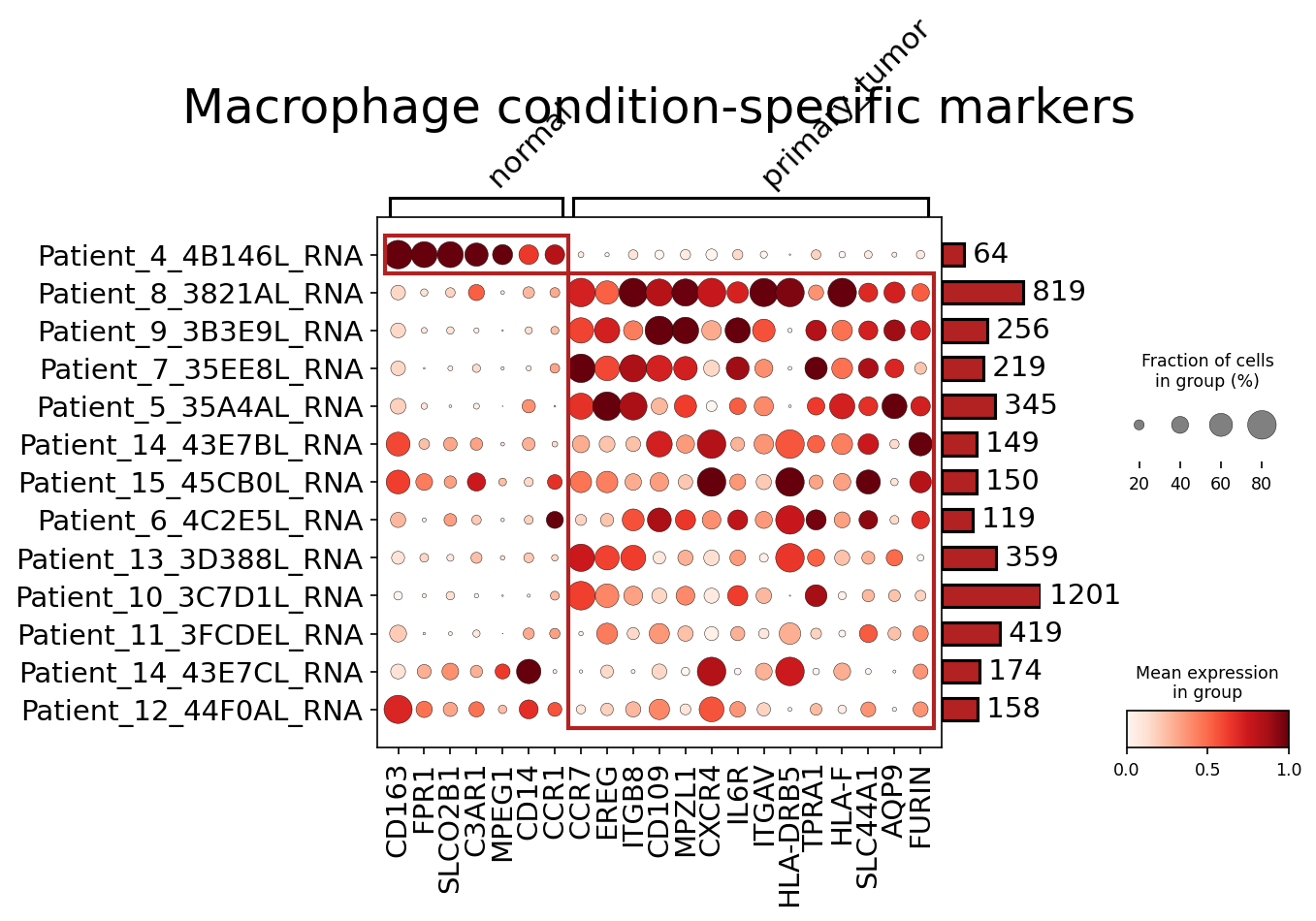
[Analysis Visualization Results]...
Analysis Overview
This analysis aimed to identify surfaceome markers that are distinctly expressed in Macrophage populations when comparing normal breast tissue macrophages to those found in primary breast tumors. By focusing on surfaceome markers, we identify proteins that are accessible on the cell surface, making them potential candidates for cell-surface-based assays (e.g., flow cytometry, immunohistochemistry) or therapeutic targeting. The plot_markers_and_expression_dot tool was used to visualize these condition-specific markers, presenting both the fraction of cells expressing each marker and its mean expression level across individual patients within each condition.
Visual Summary
The dot plot clearly differentiates Macrophage populations based on their surfaceome marker expression profiles across normal and primary tumor conditions.
- Normal Condition Markers (Left Cluster): A distinct set of surface markers, including CD163, FPR1, SLCO2B1, C3AR1, MPEG1, CD14, and CCR1, are highly expressed and prevalent in macrophages from normal breast tissue samples (e.g., Patient_4_4B146L_RNA). These markers show minimal to no expression in primary tumor macrophage populations.
- CD163 and CD14 are canonical macrophage markers, often associated with M2-like or general macrophage identity. Their enrichment in normal tissue macrophages suggests a homeostatic or resident macrophage phenotype in the healthy breast environment.
- FPR1 and C3AR1 are involved in immune and inflammatory responses.
- Primary Tumor Condition Markers (Right Cluster): A larger and more diverse cluster of surface markers is observed in macrophages derived from primary breast tumors. These include CCR7, EREG, ITGB8, CD109, MPZL1, CXCR4, IL6R, ITGAV, HLA-DRB5, TPRA1, HLA-F, SLC44A1, AQP9, and FURIN.
- These markers are consistently expressed across a majority of the primary tumor samples and show very low or absent expression in normal macrophages.
- High expression for markers like CCR7, EREG, CXCR4, IL6R, ITGAV, and HLA-DRB5 is particularly noticeable across multiple tumor patients, indicating a robust tumor-associated macrophage (TAM) signature.
The patient-specific bars on the right indicate the number of Macrophage cells analyzed per patient, showing adequate cell numbers for robust marker identification. The clustering of patients by condition (normal vs. primary tumor) visually confirms the differential expression patterns.
Biological Interpretation
The observed differential expression of surfaceome markers highlights a profound phenotypic shift in macrophages infiltrating primary breast tumors compared to those in normal breast tissue.
- Normal Macrophages: The markers identified in normal macrophages (e.g., CD163, CD14) are often associated with tissue-resident macrophages or those with a general immune surveillance and homeostatic function. While CD163 is also associated with M2-like macrophages, its prevalence in normal tissue in this context suggests it might represent a baseline or broadly defined macrophage state, distinct from the tumor-specific activation observed.
- Tumor-Associated Macrophages (TAMs): The extensive panel of surface markers upregulated in primary tumor macrophages points to a highly activated and distinct phenotype, characteristic of Tumor-Associated Macrophages (TAMs). Many of these markers are biologically significant in cancer progression:
- Chemokine Receptors (CCR7, CXCR4): Upregulation of CCR7 and CXCR4 suggests enhanced migratory capabilities and responsiveness to specific chemokines (e.g., CCL19/CCL21 for CCR7, CXCL12 for CXCR4). This is critical for TAM recruitment to the tumor microenvironment (TME) and potentially their trafficking within it or to draining lymph nodes. PubMed Search: CXCR4 macrophage cancer
- Growth Factor Signaling (EREG, IL6R): Epiregulin (EREG) is an EGFR ligand, and its expression by TAMs indicates their active participation in paracrine signaling that can promote tumor cell proliferation and survival. Upregulation of IL6R suggests TAMs are responsive to IL-6, a key pro-inflammatory cytokine in the TME, which can drive immunosuppression and tumor growth. GeneCards: EREG
- Integrins (ITGB8, ITGAV): Integrins mediate cell-extracellular matrix (ECM) and cell-cell interactions. Their upregulation suggests TAMs are actively involved in remodeling the TME, influencing cell adhesion, and potentially activating latent growth factors like TGF-β (ITGB8). UniProt: ITGB8
- Antigen Presentation & Immune Modulation (HLA-DRB5, HLA-F): While macrophages are antigen-presenting cells (APCs), altered expression of MHC class II molecules (HLA-DRB5) and non-classical MHC molecules (HLA-F) in TAMs can reflect their modified immune function within the TME, which may include altered antigen presentation or even immunosuppressive roles.
- Other markers: CD109, MPZL1, AQP9, and FURIN have diverse roles, including cell adhesion, transport, and protein processing, further highlighting the metabolic and functional adaptations of TAMs to the tumor environment.
This comprehensive signature for TAMs indicates their multifaceted roles in supporting tumor progression, angiogenesis, immune evasion, and metastasis in breast cancer.
Clinical or Translational Implications
The identification of distinct surfaceome markers for normal versus tumor-associated macrophages carries significant clinical and translational potential.
- Biomarker Discovery: The identified tumor-specific macrophage markers (e.g., CCR7, EREG, CXCR4, IL6R, ITGAV) could serve as valuable biomarkers for:
- Diagnosis/Prognosis: Assessing the infiltration and phenotype of TAMs in breast cancer biopsies using techniques like immunohistochemistry (IHC) or multiplex immunofluorescence. High expression of specific TAM markers could correlate with disease progression or prognosis.
- Monitoring Treatment Response: Changes in the expression of these markers on circulating monocytes or tissue macrophages could indicate response to therapy.
- Therapeutic Targeting: Surfaceome proteins are highly desirable targets for drug development due to their accessibility.
- Targeted Therapies: Markers like CXCR4, IL6R, and EREG, which are implicated in tumor-promoting pathways, represent potential targets for blocking TAM functions or depleting pro-tumorigenic macrophages in the TME. This could involve small molecule inhibitors or antibody-drug conjugates. For example, blocking the CXCR4-CXCL12 axis can inhibit tumor cell migration and metastasis, which might be augmented by also targeting TAMs. PubMed Search: CXCR4 inhibitor cancer therapy
- Immunotherapy: Understanding the surface markers of TAMs is crucial for designing immunotherapeutic strategies that re-educate or deplete these cells to enhance anti-tumor immunity. For instance, modifying TAMs to a more M1-like phenotype or inhibiting their immunosuppressive functions.
- Experimental Validation: The identified surface markers provide excellent candidates for further experimental validation using methods such as:
- Flow Cytometry: To precisely phenotype macrophage populations in dissociated tissue or blood.
- Immunohistochemistry (IHC) / Immunofluorescence (IF): To visualize the spatial distribution and expression levels of these markers on macrophages within tumor sections.
- Single-cell Proteomics: To confirm protein expression at the single-cell level.
By selectively targeting these surface markers, it may be possible to specifically modulate TAMs in breast cancer, thereby improving therapeutic outcomes while minimizing off-target effects on normal tissue macrophages.
17. Fibroblast Condition-Specific Surfaceome Markers in Breast Tissue
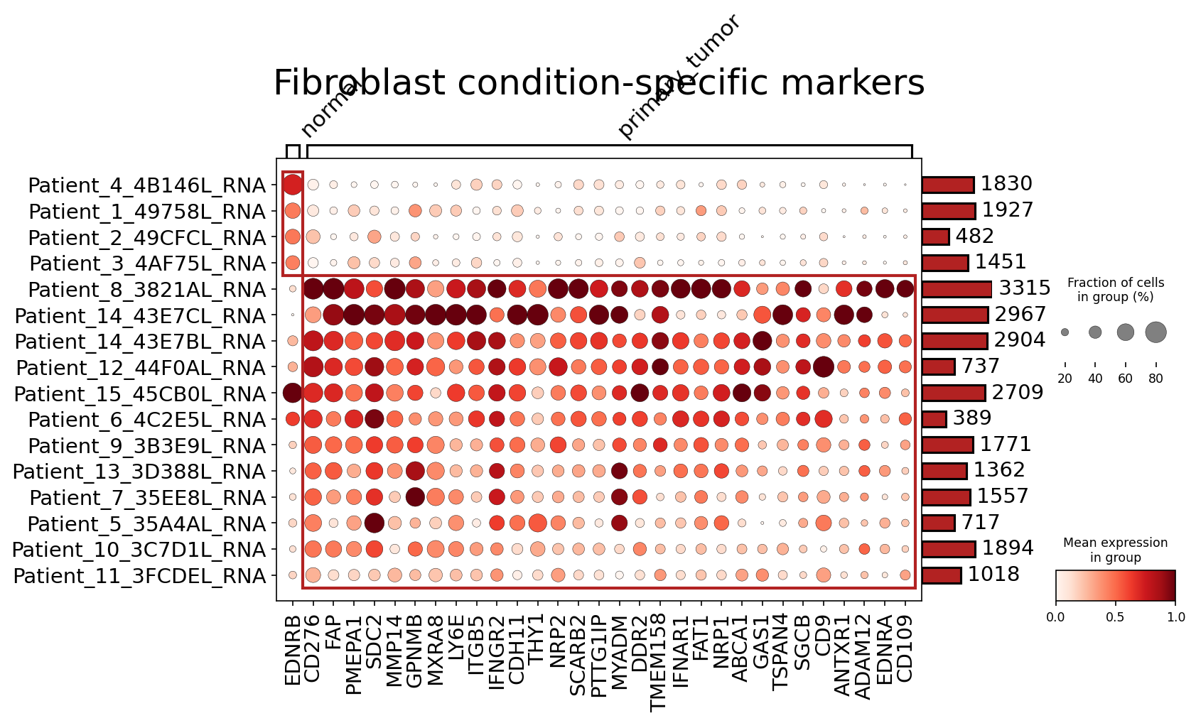
[Analysis Visualization Results]...
Analysis Overview
This analysis identifies condition-specific surfaceome markers in Fibroblast cells from breast tissue, comparing "normal" and "primary_tumor" conditions. The results are presented as a dot plot, where each row represents a patient sample, and each column represents a gene. The size of the dot indicates the fraction of cells expressing the gene, and the color intensity reflects the mean expression level within that patient's fibroblast population. Only surfaceome markers were considered, up to 50 per condition.
Visual Summary
The dot plot clearly differentiates two groups of patient samples: five "normal" samples and eleven "primary_tumor" samples, as indicated by the bar above the heatmap.
- Normal Fibroblasts: In the normal samples (top five rows), fibroblast expression of most genes displayed is generally low to moderate, with smaller dot sizes, indicating a lower fraction of cells expressing these genes. A few genes like *EDNRB*, *CD276*, and *THY1* show some intermittent expression, but without a strong, widespread pattern.
- Primary Tumor Fibroblasts: In stark contrast, the fibroblasts from primary tumor samples (bottom eleven rows, enclosed within the red boundary) exhibit a profound and consistent upregulation of numerous surfaceome markers. Many genes show uniformly high mean expression (dark red dots) and are expressed in a large fraction of cells (large dot sizes) across the majority of tumor samples. This indicates a highly activated and distinct phenotype for cancer-associated fibroblasts (CAFs).
Key upregulated markers in primary tumor fibroblasts include:
- FAP (Fibroblast Activation Protein alpha): Shows consistently high expression and widespread presence across almost all primary tumor samples.
- MMP14 (Matrix Metalloproteinase 14): Strongly expressed and prevalent in tumor samples.
- NRP1 (Neuropilin-1) and NRP2 (Neuropilin-2): Both show high expression in tumor-associated fibroblasts.
- GPNMB (Glycoprotein NMB): Highly expressed in tumor fibroblasts.
- SDC2 (Syndecan-2), ITGB5 (Integrin Beta 5), CDH11 (Cadherin-11), DDR2 (Discoidin Domain Receptor 2): These adhesion and matrix-interacting molecules are also highly expressed.
- CD276 (B7-H3), CD109, ADAM12, EDNRA: Also show significant upregulation in tumor fibroblasts.
The intensity and breadth of expression for these markers are notably higher in tumor samples compared to normal, highlighting their condition-specific nature.
Biological Interpretation
The observed upregulation of numerous surfaceome markers in fibroblasts from primary breast tumors points to a significant phenotypic shift, indicative of their transformation into Cancer-Associated Fibroblasts (CAFs). These findings align with the established role of CAFs as critical components of the tumor microenvironment (TME) in breast cancer.
- CAF Activation and ECM Remodeling: The consistent high expression of FAP [GeneCards], a well-known CAF marker, alongside MMP14 [GeneCards] and DDR2 [GeneCards], strongly suggests enhanced extracellular matrix (ECM) remodeling and degradation activities. This is crucial for creating a pro-invasive and pro-metastatic niche for tumor cells. FAP is a serine protease that remodels the ECM, and MMP14 is a membrane-bound metalloproteinase with similar functions. DDR2 is a collagen receptor that signals upon collagen binding, regulating fibroblast differentiation and matrix production.
- Cell Adhesion and Migration: Markers like ITGB5 (Integrin Beta 5) [GeneCards], SDC2 (Syndecan-2) [GeneCards], and CDH11 (Cadherin-11) are involved in cell-cell and cell-matrix interactions. Their upregulation indicates altered adhesive properties of CAFs, facilitating their migration within the TME and interaction with tumor cells and other stromal components. SDC2, a heparan sulfate proteoglycan, can bind growth factors and modulate their signaling.
- Angiogenesis and Growth Factor Signaling: The increased expression of NRP1 (Neuropilin-1) [GeneCards] and NRP2 (Neuropilin-2) [GeneCards] in tumor fibroblasts highlights their potential role in promoting angiogenesis and interacting with various growth factors (e.g., VEGF, plexins). This directly supports tumor growth and metastasis by enhancing vascularization.
- Immune Modulation: CD276 (B7-H3) [GeneCards] is an immune checkpoint molecule. Its high expression on CAFs suggests an immunosuppressive role, where fibroblasts might actively contribute to immune evasion by T cells within the tumor microenvironment.
Other Pro-tumorigenic Roles:
- GPNMB (Glycoprotein NMB) [GeneCards] is associated with tumor progression and metastasis.
- CD109 is a GPI-anchored protein involved in cell proliferation and TGF-β signaling, often linked to aggressive tumor phenotypes.
- EDNRA (Endothelin Receptor Type A) [GeneCards] is part of the endothelin system, which promotes cancer cell proliferation and survival.
This collective shift in surface protein expression underscores the multifaceted contributions of CAFs to breast cancer progression, from matrix remodeling to immune suppression and growth support.
Clinical or Translational Implications
The distinct expression of these surfaceome markers in breast cancer-associated fibroblasts offers significant clinical and translational opportunities:
- Therapeutic Targets: Several identified markers, particularly FAP, MMP14, CD276, NRP1, and NRP2, represent attractive targets for anti-cancer therapies. As surface proteins, they are readily accessible for antibody-based therapies, small molecule inhibitors, or CAR-T/NK cell strategies. Targeting CAFs directly can inhibit tumor growth, invasion, and metastasis, and may also reduce immune suppression within the TME. FAP-targeting agents are already under clinical investigation in various cancers. [PubMed Search]
- Diagnostic and Prognostic Biomarkers: The differential expression of these markers between normal and tumor fibroblasts suggests their potential utility as diagnostic or prognostic biomarkers. For instance, high levels of FAP or MMP14 in breast tissue biopsies could indicate the presence of activated CAFs, potentially correlating with disease aggressiveness or predicting patient outcomes.
- Monitoring Disease Progression and Treatment Response: Monitoring the expression of these CAF-specific markers could provide non-invasive or minimally invasive ways to track disease progression, recurrence, or the effectiveness of stromal-targeting therapies.
- Combination Therapies: Therapies targeting these CAF surface markers could be combined with existing standard-of-care treatments, such as chemotherapy, radiation, or immunotherapy, to achieve synergistic anti-tumor effects by simultaneously addressing both cancer cells and their supportive stromal niche. This approach could overcome drug resistance and enhance treatment efficacy.
18. Cell Subtype-Specific Surfaceome Marker Identification
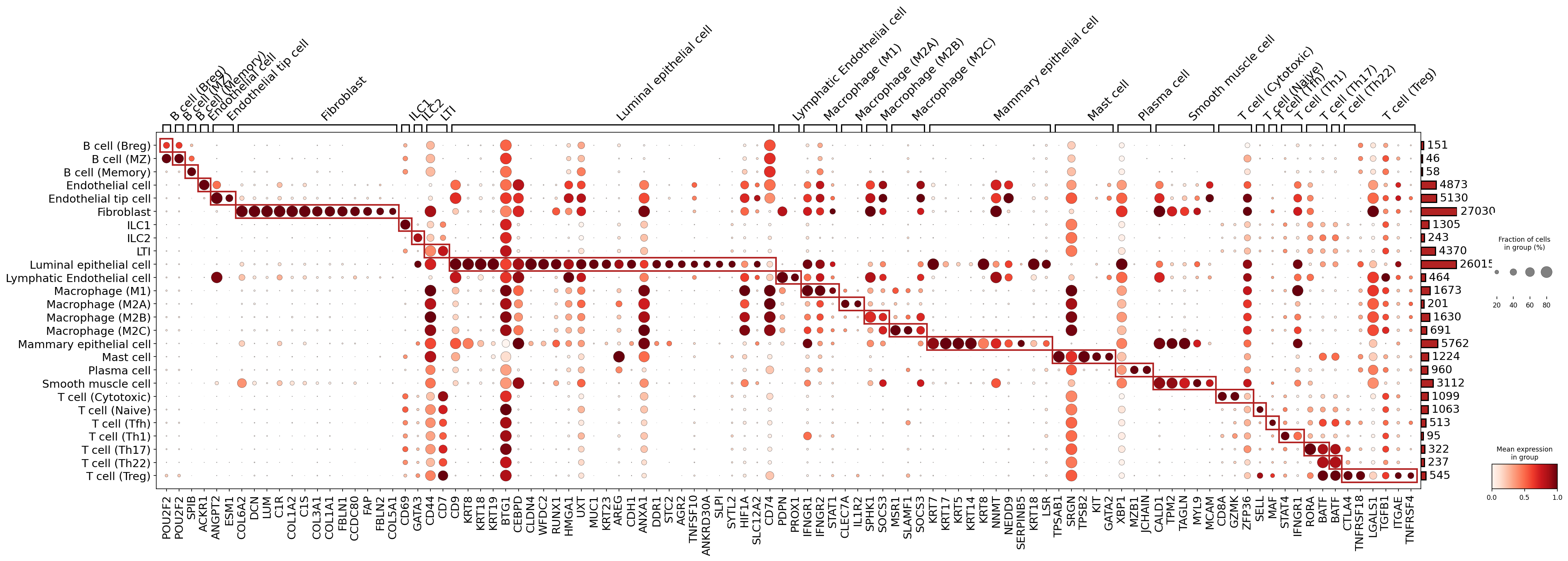
[Analysis Visualization Results]...
Analysis Overview
This analysis aimed to identify and visualize cell-type-specific surfaceome markers across various cell subsets present in the breast tissue single-cell RNA-seq dataset. The generated dot plot displays the mean expression level (color intensity) and the fraction of cells expressing a gene (dot size) for candidate markers within each celltype_subset.
It's important to note a discrepancy between the user query and the visualization. The user specifically requested "condition-specific markers for T cell CD4+". However, the provided plot shows general cell-type markers for *many different celltype_subset categories* (not just T cell CD4+), and it does not differentiate between conditions (e.g., primary_tumor vs. normal). Therefore, this interpretation will focus on the observed cell-type specificity across the displayed cell subsets, with particular attention to the CD4+ T cell subsets requested in the query, rather than condition-specific differences. While the find_cfg parameter specified surfaceome_only: True, some key intracellular transcription factors and cytokines, known to be highly specific for these cell types, are also prominently featured, indicating the strong association of these master regulators with cell identity.
Visual Summary
The dot plot effectively visualizes the expression patterns of numerous genes across 25 distinct celltype_subset categories. Each row represents a cell subset, and each column represents a gene. The red boxes highlight groups of genes that are highly specific and highly expressed within particular cell subsets.
Key observations include:
- Highly Specific Markers: Many genes exhibit high specificity (dark red, large dot) for a single cell subset, forming clear diagonal blocks. For example, POU2F2, CD79A, and CD79B are highly specific for B cell subsets, while CDH5, PECAM1, COL6A2, ANGPT2, and ESM1 are distinct for endothelial cells. Keratin genes (e.g., KRT8, KRT18, KRT19) are strong markers for epithelial cells. CD68 and MSR1 are prominent macrophage markers.
- T cell CD4+ Subsets: Focusing on the T cell CD4+ subsets (T cell (Naive), T cell (Tfh), T cell (Th1), T cell (Th17), T cell (Th22), T cell (Treg)):
- T cell (Naive): SELL (CD62L) shows strong expression and high prevalence, consistent with its role in lymphocyte homing for naive T cells.
- T cell (Tfh): Key markers include BCL6 (a master regulator transcription factor) and PDCD1 (PD-1, an immune checkpoint receptor), reflecting their critical roles in B cell help and germinal center reactions. TNFRSF4 (OX40) is also a strong surface marker.
- T cell (Th1): STAT4 (a transcription factor) and IFNG (IFN-gamma, a signature cytokine) are prominently shown, consistent with their pro-inflammatory roles.
- T cell (Th17): RORC (RORγt, a master regulator transcription factor) and BATF (a transcription factor) are strong markers for this subset, which is involved in host defense and autoimmune responses.
- T cell (Treg): FOXP3 (a master regulatory transcription factor) and the surface receptors CTLA4 and TNFRSF18 (GITR) are clearly identified, underscoring their immunosuppressive functions.
- Other T cell Subsets: T cell (Cytotoxic) (likely CD8+ T cells) are characterized by CD8A and granzymes like GZMK, highlighting their effector function.
- Gene Expression Patterns: The dot size and color provide insights into the homogeneity and expression levels of markers within each group. Larger, darker red dots indicate highly expressed genes present in a high fraction of cells, signifying robust markers.
Biological Interpretation
The identified markers provide valuable insights into the identity and functional characteristics of various immune and stromal cell populations within the breast tissue. For T cell CD4+ subsets, these markers are critical for their differentiation, function, and interaction with other cells in the tumor microenvironment.
- T cell (Naive): The presence of SELL (CD62L) confirms their naive state, enabling them to recirculate through lymphoid organs. [GeneCards]
- T cell (Tfh): Markers like PDCD1 (PD-1) and TNFRSF4 (OX40) are integral to Tfh cell function, mediating interactions with B cells in germinal centers and promoting antibody production. BCL6 is the master transcription factor for Tfh development. [GeneCards] [GeneCards]
- T cell (Th1): The expression of STAT4 and IFNG reflects their role in cell-mediated immunity, particularly against intracellular pathogens and tumors, often promoting a pro-inflammatory environment. [GeneCards]
- T cell (Th17): RORC and BATF are key for the development of Th17 cells, which produce IL-17 and contribute to inflammatory responses and defense against extracellular pathogens. [GeneCards]
- T cell (Treg): FOXP3, CTLA4, and TNFRSF18 (GITR) are hallmarks of regulatory T cells, which suppress immune responses and maintain peripheral tolerance. In cancer, Tregs often contribute to immune evasion. [GeneCards] [GeneCards]
The identification of these cell-type-specific markers, including both surface proteins and master transcription factors, provides a comprehensive molecular signature for each cell subset. The inclusion of intracellular markers despite the surfaceome_only setting highlights their critical role in defining cell identity, suggesting that for certain cell types, intracellular master regulators are indispensable for accurate identification and functional understanding.
Clinical or Translational Implications
Although this analysis did not provide *condition-specific* markers for T cell CD4+ as initially requested, the identified cell-type-specific surfaceome markers are highly valuable for several translational applications:
- Cell Isolation and Characterization: Surface markers like SELL, PDCD1, CTLA4, and TNFRSF4 can be used for flow cytometry-based immunophenotyping or cell sorting to isolate specific T cell CD4+ subsets from complex breast tissue samples. This enables further functional studies of these populations.
- Biomarker Discovery: These markers can serve as potential diagnostic or prognostic biomarkers. For instance, the infiltration levels of specific T cell CD4+ subsets (e.g., Treg or Tfh) identified by their unique surface markers could correlate with disease progression or response to therapy in breast cancer.
- Therapeutic Targets: Surface receptors such as PDCD1 (PD-1) on Tfh cells and CTLA4 or TNFRSF18 (GITR) on Treg cells are established or emerging targets for immunomodulatory therapies, particularly in cancer.
- Targeting PDCD1 (e.g., with anti-PD-1 antibodies) can reinvigorate exhausted T cells and is a cornerstone of current cancer immunotherapy. [PubMed Search]
- Modulating CTLA4 activity (e.g., with ipilimumab) also serves to enhance anti-tumor immunity by inhibiting Treg function or enhancing effector T cell activation. [PubMed Search]
- TNFRSF18 (GITR) agonists are being explored as a means to deplete Tregs or activate effector T cells, representing a promising avenue for novel immunotherapies. [PubMed Search]
Further analysis to identify *condition-specific* markers within T cell CD4+ subsets would directly inform how these populations are altered in primary tumors compared to normal tissue, leading to more targeted therapeutic strategies or prognostic indicators.
19. Epithelial Cell Cycle Gene YWHAZ Upregulation in Primary Breast Tumors
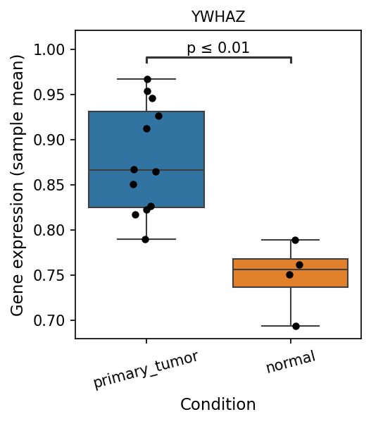
[Analysis Visualization Results]...
Analysis Overview
This analysis investigates the differential expression of genes related to the Cell Cycle pathway in Epithelial cells (the tumor-origin cell type) from breast tissue. Specifically, a box plot displays the expression levels of the gene YWHAZ, comparing primary tumor samples to normal samples, highlighting statistically significant differences. The gene expression values represent the mean expression per sample.
Visual Summary
The box plot shows the expression of YWHAZ in Epithelial cells across two conditions: 'primary_tumor' and 'normal'.
- Differential Expression: YWHAZ gene expression is significantly higher in primary tumor samples compared to normal samples (p ≤ 0.01).
- Expression Levels: The median expression of YWHAZ in primary tumor Epithelial cells is approximately 0.86, with a considerable spread of sample means ranging from about 0.79 to 0.97. In contrast, normal Epithelial cells show a lower median expression of approximately 0.75, with a tighter distribution of sample means between roughly 0.69 and 0.79.
- Variability: While both conditions show some variability, the 'primary_tumor' group exhibits a wider interquartile range and a broader range of individual sample means, suggesting more heterogeneity in YWHAZ expression within tumor samples compared to normal samples.
Biological Interpretation
YWHAZ (Tyrosine 3-monooxygenase/tryptophan 5-monooxygenase activation protein, zeta polypeptide), also known as 14-3-3 zeta, is a crucial member of the 14-3-3 protein family. These proteins are highly conserved regulatory molecules that modulate the function of various client proteins involved in signal transduction, cell cycle progression, apoptosis, and cellular metabolism through protein-protein interactions [1].
The observed significant upregulation of YWHAZ in Epithelial cells from primary breast tumors, compared to normal breast tissue, is biologically relevant given its established role in cell cycle regulation and cancer biology.
- Cell Cycle Progression: As part of the gene list for "Cell Cycle pathway-related genes," the increased expression of YWHAZ suggests its potential involvement in driving the uncontrolled proliferation characteristic of tumor cells. 14-3-3 proteins, including YWHAZ, can regulate key cell cycle checkpoints and progression by interacting with cyclins, CDKs, and cell cycle inhibitors [2].
- Oncogenic Role: In various cancers, including breast cancer, YWHAZ has been reported to be overexpressed and often functions as an oncogene. Its overexpression can promote cell growth, inhibit apoptosis, enhance cell invasion, and contribute to chemoresistance [3]. This aligns with its elevated expression in tumor-origin epithelial cells.
- Breast Cancer Context: Given that Epithelial cells are the origin of breast cancer, the specific upregulation of YWHAZ in these malignant cells points towards its potential contribution to breast tumor initiation and progression within the breast microenvironment.
Clinical or Translational Implications
The differential expression of YWHAZ in breast cancer Epithelial cells suggests several potential clinical and translational implications:
- Biomarker Potential: YWHAZ could serve as a potential diagnostic or prognostic biomarker for breast cancer. Elevated YWHAZ expression in tumor biopsies or circulating tumor cells might indicate tumor presence or aggressiveness [4].
- Therapeutic Target: Given its oncogenic role and involvement in key cellular processes like the cell cycle, YWHAZ could represent a promising therapeutic target in breast cancer. Inhibiting YWHAZ or its interactions might impede tumor growth and progression, potentially sensitizing cancer cells to existing therapies.
---
References:
- General 14-3-3 function: Yaffe, M. B. (2002). 14-3-3 proteins: universal regulators of signalling. *Current Opinion in Cell Biology*, *14*(2), 209-216. PubMed search: 14-3-3 proteins cell cycle regulation
- 14-3-3 proteins in cell cycle: Obsil, T., & Obsilova, V. (2008). 14-3-3 proteins: a family of versatile regulators of cell cycle progression. *Vitamins and Hormones*, *78*, 27-57. PubMed search: 14-3-3 proteins cell cycle
- YWHAZ in cancer: Wang, W., et al. (2018). The role of 14-3-3ζ in human cancer. *Molecular Cancer*, *17*(1), 1-13. PubMed search: YWHAZ oncogene breast cancer
- YWHAZ as biomarker in breast cancer: GeneCards entry for YWHAZ, often includes links to disease associations and biomarker studies. GeneCards: YWHAZ
20. Epithelial Cell Gene Ontology Analysis: Differential Pathway Enrichment Across Ploidy and Tumor Status in Breast Tissue
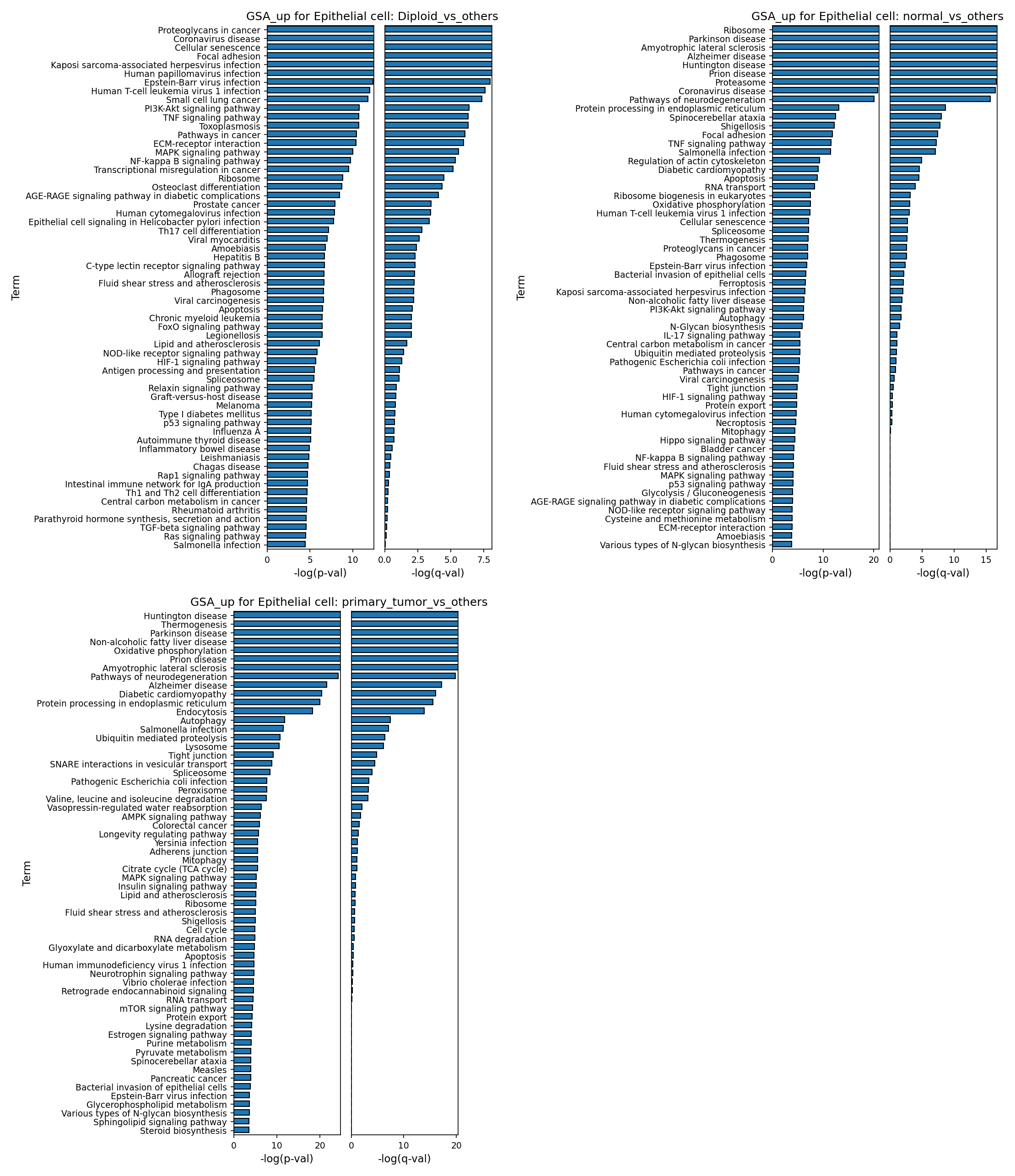
[Analysis Visualization Results]...
Analysis Overview
This analysis utilizes Gene Ontology (GSA) to identify enriched biological pathways in epithelial cells from breast tissue. The results are presented as bar plots, showing the negative logarithm of p-values (-log(p-val)) and q-values (-log(q-val)) for pathway enrichment across three key comparisons:
- Diploid epithelial cells vs. Aneuploid epithelial cells: Highlighting pathways associated with ploidy status within epithelial cells.
- Normal epithelial cells vs. Primary tumor epithelial cells: Identifying pathways characteristic of the healthy epithelial state.
- Primary tumor epithelial cells vs. Normal epithelial cells: Revealing pathways enriched in cancerous epithelial cells.
The goal is to provide biological insights into the functional shifts occurring in epithelial cells in the context of breast cancer progression and ploidy changes.
Visual Summary
The bar plots display the top 60 enriched GO terms, ranked by -log(p-val), for each comparison. Higher bars indicate more significant enrichment.
- Diploid_vs_others (likely Diploid vs Aneuploid epithelial cells): This plot shows strong enrichment for pathways related to cancer (e.g., "Proteoglycans in cancer," "Pathways in cancer"), cell adhesion ("Focal adhesion," "ECM-receptor interaction"), and key signaling cascades (e.g., "PI3K-Akt signaling pathway," "MAPK signaling pathway," "NF-kappa B signaling pathway"). "Cellular senescence" is also highly enriched.
- normal_vs_others (likely Normal vs Primary tumor epithelial cells): This plot is dominated by terms related to fundamental cellular machinery and homeostasis. "Ribosome," "Proteasome," "Protein processing in endoplasmic reticulum," "Oxidative phosphorylation," and "Apoptosis" are prominently enriched. "Cellular senescence" and "Focal adhesion" also appear. A notable presence of neurodegenerative disease terms is observed across multiple comparisons.
- primary_tumor_vs_others (likely Primary tumor vs Normal epithelial cells): This plot reveals significant enrichment in metabolic processes ("Oxidative phosphorylation," "Autophagy," "Ubiquitin mediated proteolysis," "Lysosome," "Citrate cycle"), cell cycle ("Cell cycle"), and altered cell-cell junctions ("Tight junction," "Adherens junction"). Signaling pathways like "AMPK signaling pathway" and "MAPK signaling pathway" are also enriched.
Biological Interpretation
Functional Signatures of Diploid vs. Aneuploid Epithelial Cells
In the breast tissue epithelial compartment, diploid cells show upregulation of pathways associated with early carcinogenesis, cell-matrix interactions, and tumor suppression when compared to aneuploid cells. The enrichment of "Proteoglycans in cancer" and "Pathways in cancer" suggests an active role within the tumor microenvironment, possibly reflecting either less transformed cells or those responding to oncogenic stimuli in a structured manner. The prominence of "Focal adhesion" and "ECM-receptor interaction" indicates intact or heightened communication with the extracellular matrix, critical for maintaining tissue architecture or initiating remodeling. "Cellular senescence," a known tumor-suppressive mechanism that limits the proliferative capacity of damaged cells, is also enriched, suggesting that diploid cells may retain a greater capacity for cell cycle arrest in response to stress. The enrichment of viral infection pathways (e.g., HPV, EBV) might reflect a general activation of innate immune or stress response pathways that are not necessarily indicative of direct viral infection in breast cancer, but rather common cellular responses to perturbation.
Homeostatic Processes in Normal Epithelial Cells
Normal breast epithelial cells, when compared to their malignant counterparts, exhibit a robust enrichment in pathways central to cellular anabolism, catabolism, energy production, and programmed cell death. High enrichment of "Ribosome," "Proteasome," and "Protein processing in endoplasmic reticulum" underscores their active role in protein synthesis, folding, and degradation, essential for maintaining cellular function and integrity. "Oxidative phosphorylation" highlights efficient energy metabolism. The upregulation of "Apoptosis" and "Cellular senescence" pathways emphasizes the tightly regulated cell turnover and damage control mechanisms inherent to healthy tissue, crucial for preventing aberrant cell accumulation. "Focal adhesion" and "Regulation of actin cytoskeleton" reflect the organized structural and mechanical properties of healthy epithelial layers.
Oncogenic Reprogramming in Primary Tumor Epithelial Cells
Primary tumor epithelial cells display a clear reprogramming of metabolism, altered cell cycle control, and changes in cell junction dynamics when compared to normal epithelial cells. The enrichment of "Cell cycle" pathways is a direct hallmark of uncontrolled proliferation, a defining feature of cancer. Metabolic shifts are evident with increased "Oxidative phosphorylation," "Autophagy," "Ubiquitin mediated proteolysis," and activation of the "Citrate cycle" (TCA cycle), indicating altered energy demands and nutrient utilization often associated with the Warburg effect and other cancer-specific metabolic adaptations. Dysregulation of cell-cell adhesion, as suggested by changes in "Tight junction" and "Adherens junction" pathways, is critical for tumor progression, enabling cells to detach from the primary tumor and potentially invade surrounding tissues. Activation of "AMPK signaling pathway" and "MAPK signaling pathway" further points to dysregulated growth and survival signals. The consistent appearance of pathways related to neurodegenerative diseases (e.g., Huntington, Parkinson) across different comparisons might indicate shared underlying mechanisms of cellular stress, protein misfolding, or metabolic dysfunction that become perturbed in various pathological states, including cancer, rather than a direct etiological link to neurodegeneration in breast cancer.
Clinical or Translational Implications
The distinct pathway enrichments in diploid vs. aneuploid and normal vs. primary tumor epithelial cells offer potential avenues for clinical translation:
- Targeting Metabolic Vulnerabilities: The prominent metabolic reprogramming in primary tumor epithelial cells (e.g., oxidative phosphorylation, autophagy, TCA cycle) suggests that specific metabolic enzymes or pathways could be therapeutic targets to selectively inhibit cancer cell growth, potentially starving tumor cells or sensitizing them to other therapies.
- Cell Cycle and Cell Adhesion as Therapeutic Targets: The clear upregulation of cell cycle pathways in tumor cells reinforces their role as primary targets for cytotoxic and cell cycle-specific drugs. Furthermore, alterations in "Tight junction" and "Adherens junction" pathways could be exploited to inhibit tumor invasion and metastasis, perhaps by restoring normal cell-cell contacts or disrupting tumor-specific adhesion mechanisms.
- Ploidy as a Prognostic Marker: The distinct biological signatures of diploid versus aneuploid epithelial cells might contribute to understanding tumor heterogeneity and could inform prognostic stratification or treatment selection. Pathways enriched in diploid cells, such as "Cellular senescence," might indicate a subpopulation with a different response to therapy or a slower progression rate.
- Understanding Tumor Microenvironment: The involvement of "Proteoglycans in cancer" and "ECM-receptor interaction" in diploid cells highlights the importance of the extracellular matrix in modulating tumor behavior, suggesting that therapies targeting matrix components or cell-matrix interactions could be beneficial.
Understanding these functional shifts at the pathway level provides a comprehensive view of how breast epithelial cells transform from a healthy state to a malignant one, with distinct features also observed based on their ploidy status. This knowledge can help in identifying novel therapeutic strategies and biomarkers for breast cancer.
21. Gene Set Enrichment Analysis (GSEA) in Breast Cancer Endothelial and Epithelial Cells
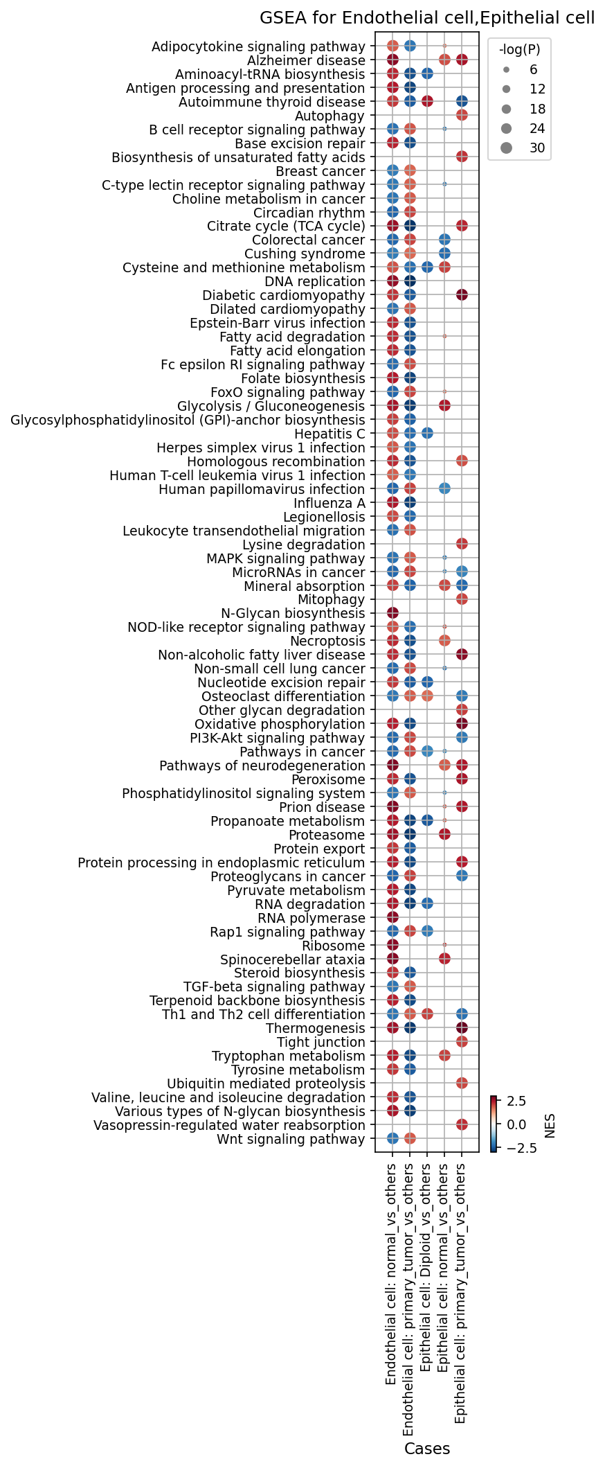
[Analysis Visualization Results]...
Analysis Overview
This analysis presents Gene Set Enrichment Analysis (GSEA) results, visualized as a dot plot, for two key cell types: Endothelial cells and Epithelial cells. GSEA identifies biological pathways or gene sets that are coordinately up- or down-regulated in a given condition compared to a reference or 'other' conditions. Here, we investigate pathway enrichment in Endothelial cells from normal tissue versus other conditions, and from primary tumor tissue versus other conditions. For Epithelial cells, we compare diploid cells versus other epithelial cells, and primary tumor epithelial cells versus other epithelial cells. The dot size represents the statistical significance (-log(p-value)), and the color indicates the Normalized Enrichment Score (NES), where red signifies positive enrichment (pathway genes generally upregulated) and blue signifies negative enrichment (pathway genes generally downregulated).
Visual Summary
The dot plot effectively illustrates the differential enrichment of a wide range of biological pathways across the analyzed cell types and conditions.
- Endothelial cells: In normal tissue, Endothelial cells generally show negative enrichment (blue dots) for pathways associated with cell cycle, DNA replication, and various "Pathways in cancer" when compared to other endothelial cells. Conversely, in primary tumor tissue, Endothelial cells exhibit strong positive enrichment (large red dots) for a broad spectrum of proliferation-associated and cancer-driving pathways.
- Epithelial cells: Diploid Epithelial cells, when compared to other epithelial cells (likely including aneuploid/tumor cells), show significant negative enrichment (large blue dots) for many proliferative and cancer-related pathways. In stark contrast, primary tumor Epithelial cells display an extensive and highly significant positive enrichment (very large, dark red dots) across nearly all evaluated cancer-related, proliferative, and metabolic pathways.
- Commonalities and Differences: Pathways related to DNA replication, cell cycle, MAPK, PI3K-Akt signaling, and "Pathways in cancer" are consistently and strongly positively enriched in both primary tumor Endothelial and Epithelial cells. Metabolic pathways such as "Glycolysis / Gluconeogenesis" and "Fatty acid degradation" show more nuanced and condition-specific enrichment patterns.
Biological Interpretation
The GSEA results reveal profound shifts in cellular biology associated with breast cancer progression in both the cancerous epithelial cells and the surrounding tumor microenvironment (specifically, endothelial cells).
Tumor-Associated Proliferation and Metabolism:
- Endothelial cells in primary tumor show a dramatic upregulation of pathways critical for angiogenesis and tumor growth, including DNA replication, Homologous recombination, Cell cycle, MAPK signaling pathway, PI3K-Akt signaling pathway, and various "Pathways in cancer" [NCBI]. This indicates active proliferation of endothelial cells to form new blood vessels that supply the growing tumor, a hallmark of cancer.
- Epithelial cells in primary tumor exhibit an even more widespread and intense upregulation of similar proliferative pathways. Additionally, pathways like Wnt signaling pathway [GeneCards], TGF-beta signaling pathway, Protein processing in endoplasmic reticulum, and Ubiquitin mediated proteolysis are highly enriched. This points to uncontrolled cell division, altered cell-cell communication, and active protein synthesis and degradation machinery supporting rapid tumor growth and adaptation. Metabolic pathways such as Glycolysis / Gluconeogenesis are also highly active, consistent with the "Warburg effect" observed in many cancers, where cells rely on glycolysis even in the presence of oxygen [PubMed Search].
Quiescence in Normal/Diploid Cells:
- Normal Endothelial cells show a general suppression (negative NES) of proliferation-related pathways, suggesting a quiescent state compared to their tumor-associated counterparts. This highlights the activation of endothelial cells within the tumor microenvironment.
- Diploid Epithelial cells also display a marked downregulation of proliferation and cancer-associated pathways when compared to 'others' (presumably including aneuploid/tumor cells). This provides a clear contrast, indicating that non-malignant, genetically stable epithelial cells maintain tightly controlled growth. Their positive enrichment for Glycolysis / Gluconeogenesis and Oxidative phosphorylation may reflect specific metabolic adaptations in non-tumorigenic contexts.
Signaling and Stress Responses:
- Pathways like HIF-1 signaling pathway, Protein processing in endoplasmic reticulum, and Ribosome are notably upregulated in primary tumor epithelial cells, reflecting cellular responses to hypoxia, nutrient deprivation, and increased protein synthesis demands characteristic of the tumor microenvironment.
- The enrichment of viral infection pathways (e.g., Herpes simplex virus 1 infection, Human papillomavirus infection) might represent immune evasion mechanisms or indirect effects within the tumor microenvironment, although their direct causal role here would require further investigation.
Clinical or Translational Implications
The distinct pathway enrichments observed in primary tumor cells, both epithelial and endothelial, hold significant clinical and translational relevance for breast cancer:
- Therapeutic Targets: The consistently upregulated pathways in tumor-associated cells, such as PI3K-Akt, MAPK, and Wnt signaling, represent established or emerging therapeutic targets in breast cancer [PubMed Search]. Targeting these pathways could inhibit tumor proliferation, survival, and angiogenesis.
- Metabolic Reprogramming: The strong enrichment of glycolysis and other metabolic pathways in tumor cells suggests that targeting tumor metabolism could be an effective anti-cancer strategy. Inhibitors of key glycolytic enzymes are under investigation for their potential to starve cancer cells [PubMed Search].
- Angiogenesis Inhibition: The activation of proliferative pathways in tumor Endothelial cells underscores the importance of angiogenesis in breast cancer progression. Anti-angiogenic therapies, which aim to inhibit the formation of new blood vessels, are a recognized treatment approach to limit tumor growth and metastasis [PubMed Search].
- Biomarker Discovery: The specific pathway signatures could serve as biomarkers for identifying tumor cells, assessing tumor aggressiveness, or monitoring treatment response.
22. Discussion
The comprehensive single-cell analysis of breast tissue reveals a dramatic reprogramming of the cellular landscape and communication networks in primary tumors, fundamentally distinct from normal tissue homeostasis. The identification of aneuploid epithelial cells, which strongly co-localize with primary tumor regions and exhibit extensive copy number variations, unequivocally marks them as the malignant population. These tumor-origin cells display upregulated surface markers such as EGFR, MUC1, ERBB3, and CA12, along with significant activation of cell cycle pathways, notably YWHAZ. Gene Ontology and GSEA further highlight their metabolic reprogramming, favoring glycolysis and altered oxidative phosphorylation, alongside activated proliferative pathways like PI3K-Akt, MAPK, and Wnt signaling, all crucial for uncontrolled tumor growth.
The tumor microenvironment (TME) undergoes extensive remodeling. Fibroblasts transform into Cancer-Associated Fibroblasts (CAFs), characterized by high expression of FAP, MMP14, NRP1/2, CD276, and CDH11. These CAFs are central to pathological extracellular matrix (ECM) remodeling, mediating extensive integrin-collagen and fibronectin-integrin interactions that contribute to tissue stiffness and support tumor invasion. Macrophages, or Tumor-Associated Macrophages (TAMs), adopt a distinct phenotype in tumors, upregulating surface markers like CCR7, CXCR4, EREG, IL6R, and ITGAV. Their engagement in cell-cell interactions, particularly via the MIF-CD74/CXCR4 axis and SPP1-CD44/integrin signaling, underscores their pro-inflammatory, immunosuppressive, and pro-angiogenic roles. The immune cell compartment also shifts, with primary tumors showing increased proportions of immunosuppressive Tregs, pro-inflammatory Th17 cells, and T follicular helper (Tfh) cells, while innate lymphoid cells like LTI and ILC1 are often diminished, indicative of an altered and often suppressive anti-tumor immune response. Endothelial cells within tumors exhibit a highly proliferative and pro-angiogenic signature, with strong VEGFA-VEGFR signaling, essential for tumor vascularization. Cross-talk involving EGFR and TGF-beta pathways is intensified and broadened across multiple cell types in the TME, highlighting pervasive growth factor and immunomodulatory signaling that fuels tumor progression. The specific activation of TGFB1-integrin_avb6_complex interactions in tumor suggests a unique mechanism of TGF-beta activation driving pro-tumorigenic effects.
Hypotheses:
- Aneuploid epithelial cells, marked by specific surfaceome and cell cycle gene upregulation, are the primary drivers of tumor growth and actively orchestrate the remodeling of the surrounding microenvironment in breast cancer.
- The phenotypic shift of fibroblasts to CAFs and macrophages to TAMs in the breast tumor microenvironment is critical for establishing an immunosuppressive and pro-invasive niche, directly contributing to disease progression.
- Dysregulated cell-cell communication, particularly through specific ligand-receptor axes like MIF-CD74, SPP1-integrin, and enhanced growth factor signaling (EGFR, TGF-beta), dictates the malignant behavior of tumor cells and their interactions with stromal and immune components.
- The observed metabolic reprogramming in primary tumor epithelial cells represents a key vulnerability that can be exploited for targeted therapeutic interventions.
Potential therapeutic targets:
- EGFR/ERBB3 (HER3): Overexpressed on tumor-origin epithelial cells and activated in tumor-specific cell-cell interactions, driving cell proliferation, survival, and migration. Evidence: Upregulated in primary tumor epithelial cells (surfaceome markers, Image 19). EGFR ligands (AREG, HBEGF, EREG) show enhanced interaction in tumor CCIs (Image 13, Image 16). GSEA shows positive enrichment of MAPK and PI3K-Akt signaling pathways in tumor epithelial cells (Image 27). Validation: Test existing EGFR/HER3 inhibitors (e.g., gefitinib, erlotinib, antibody therapies) in breast cancer patient-derived organoids or xenograft models. Evaluate treatment response via tumor growth inhibition, cell viability, and changes in downstream signaling pathways.
- FAP (Fibroblast Activation Protein alpha): Highly specific marker for Cancer-Associated Fibroblasts (CAFs), involved in ECM remodeling and creating a pro-tumorigenic, immunosuppressive microenvironment. Evidence: Robustly and consistently upregulated in primary tumor fibroblasts (surfaceome markers, Image 21). Involved in extensive collagen-integrin and fibronectin-integrin interactions (Image 14, Image 18). Validation: Assess the efficacy of FAP-targeting agents (e.g., FAP-specific antibodies, FAP-targeting ADCs) in preclinical breast cancer models. Monitor changes in tumor stiffness, ECM composition, and immune cell infiltration.
- CXCR4 / CXCL12 axis: Upregulated in tumor-associated macrophages (TAMs) and involved in fibroblast-endothelial cell interactions, promoting TAM recruitment, angiogenesis, and tumor metastasis. Evidence: CXCR4 is a highly upregulated surface marker in primary tumor macrophages (Image 20). CXCL14-CXCR4 interactions are prominent between fibroblasts and endothelial cells in primary tumors (Image 18). Validation: Utilize CXCR4 antagonists (e.g., plerixafor) in combination therapies in vivo to assess impact on TAM infiltration, angiogenesis, tumor growth, and metastatic potential. Conduct in vitro migration assays for TAMs and endothelial cells.
- MIF (Macrophage Migration Inhibitory Factor) / CD74 axis: Highly active in macrophage-driven inflammation, immunosuppression, and tumor progression within the tumor microenvironment. Evidence: MIF-CD74/CXCR4 interactions are highly active across multiple macrophage-involved cell pairs (Mac/Mac, Mac/Fib, Mac/Diploid Epi, Mac/Aneuploid Epi) in primary tumors (Image 13). Validation: Test small molecule inhibitors or blocking antibodies targeting MIF or CD74 in preclinical models. Evaluate their impact on TAM polarization, immune evasion, and tumor growth, potentially in combination with immunotherapies.
- TGF-beta pathway (specifically TGFB1-integrin_avb6_complex): Intensified and dysregulated signaling in the tumor microenvironment, promoting fibrosis, immune suppression, EMT, and activating latent TGF-beta. Evidence: TGF-beta signaling is broadly active in tumor CCIs (Image 13, Image 16). The TGFB1-integrin_avb6_complex interaction is specifically observed in the primary tumor context, involving aneuploid epithelial cells and fibroblasts (Image 16). Validation: Target integrin αvβ6 specifically with blocking antibodies or use pan-TGF-beta inhibitors. Assess their effect on ECM deposition, tumor cell invasion, and immune cell function in vitro and in vivo. Measure activation of TGF-beta downstream targets.
Follow-up validation ideas:
- Perform spatial transcriptomics or multiplex immunofluorescence to confirm the localization and spatial interactions of aneuploid epithelial cells, CAFs (FAP+, MMP14+), and TAMs (CXCR4+, EREG+) within the tumor microenvironment.
- Conduct in vitro co-culture experiments using isolated aneuploid epithelial cells, CAFs, and TAMs to validate the functional impact of specific ligand-receptor interactions (e.g., MIF-CD74, SPP1-CD44) on cell proliferation, migration, and immune modulation using blocking antibodies or gene editing.
- Utilize patient-derived organoid or xenograft models to test the efficacy of inhibitors targeting key dysregulated pathways (e.g., EGFR, TGF-beta, CXCR4, FAP) or their combination on tumor growth, metastasis, and TME composition.
- Validate the prognostic and predictive value of identified surface markers (e.g., EGFR, MUC1, FAP, CXCR4) and altered immune cell proportions (Tregs, Th17) in larger independent breast cancer cohorts using immunohistochemistry and flow cytometry.
- Employ targeted metabolomics to confirm the metabolic shifts (e.g., glycolysis, oxidative phosphorylation) in isolated primary tumor epithelial cells and assess the impact of metabolic inhibitors on their viability and proliferation.
Limitations:
This report is based on single-cell RNA sequencing data, which provides high-resolution cellular insights but has inherent limitations. Observed gene expression patterns and inferred cell-cell interactions are correlative and require functional validation to establish causality. Copy number variation inference from RNA-seq is an approximation and may not capture all genomic alterations. While the UMAP visualization provides a projection of cellular states, it does not directly represent the spatial organization of cells, which is crucial for contextualizing cell-cell interactions. The findings are derived from a specific cohort of breast cancer patients, and results may vary in broader populations or different breast cancer subtypes. Further in vitro and in vivo experimental validation is necessary to confirm the functional relevance of the identified markers and pathways.
23. Query List
- Show UMAP colored by condition, sample, celltype_major, celltype_minor, ploidy_dec, and celltype_subset in 2 columns, and save it.
- Show major celltype scores on UMAP and save them.
- Show the overall Celltype_subset marker expression dot plot and save it. Keep target_cell as None and keep the other arguments at their default values.
- For tumor-origin cells and unassigned cells, show a CNV heatmap grouped by sample, along with a summary of significantly amplified copy number regions, and save it.
- Show CNV patterns on UMAP, including major celltype, minor celltype, ploidy_dec, condition, and sample in 2 columns, and save it.
- Show a population bar plot for minor cell types and save it.
- Show a subset population bar plot for T cells and save it.
- For T cell subsets, show a box plot for populations that show significant differences between conditions and save it. Set ncols appropriately based on the total number of panels.
- Show a subset population bar plot for macrophages and save it.
- For tumor-origin cells and unassigned cells, show a ploidy population bar plot and save it.
- Show cell-cell interaction patterns for tumor-origin cells, fibroblasts, macrophages, and T cells, by condition and save them. Select up to 80 cell-cell interactions per condition.
- Show cell-cell interactions by condition and save the result. Select at most 80 cell-cell interactions for each condition.
- For genes related to immune checkpoint and cell cycle pathways, show cell-cell interactions and save them.
- For major immune and stromal cells, find statistically significant differences in cell-cell interactions between conditions and show them as a dot plot, and save it. Set max_n_items_per_group to 25.
- Show the condition-specific markers for tumor-origin cells (Epithelial cell) as a dot plot and save the result. Use only surfaceome markers, up to 50 markers per condition.
- For Macrophage, extract condition-specific markers and show them as a dot plot, and save it. Include only surfaceome markers, up to 50 per condition.
- For Fibroblast, extract condition-specific markers and show them as a dot plot, and save it. Include only surfaceome markers, up to 50 per condition.
- For T cell CD4+, extract condition-specific markers and show them as a dot plot, and save it. Include only surfaceome markers, up to 50 per condition.
- For Epithelial cell (the tumor-origin cell type), show a box plot of Cell Cycle pathway-related genes with statistically significant expression differences between conditions, and save it. Set max_n_items_to_plot to 24, and ncols such that the panel ratio is approximately 2x3.
- Show Gene Ontology (GSA) analysis results for epithelial cells as bar plots and save the result.
- Show Gene Set Enrichment Analysis (GSEA) results for major cell types as a dot plot and save it. Use 'RdBu_r' for the colormap and set n_pws_to_show to 80.